ناقش قسم علوم الحياة في كلية التربية للعلوم الصرفة (ابن الهيثم) اطروحة الدكتوراه الموسومة (دراسة شكليائية، نسجية ونسجية قياسية لتكوين الكبد في جنين الدجاج المحلي Gallus gallus domesticus  (1758 Linnaeus) ) للطالبة (داليا حسن ظاهر علي) التي انجزتها باشراف التدريسية في القسم (أ.د. نهلة عبد الرضا البكري ) .

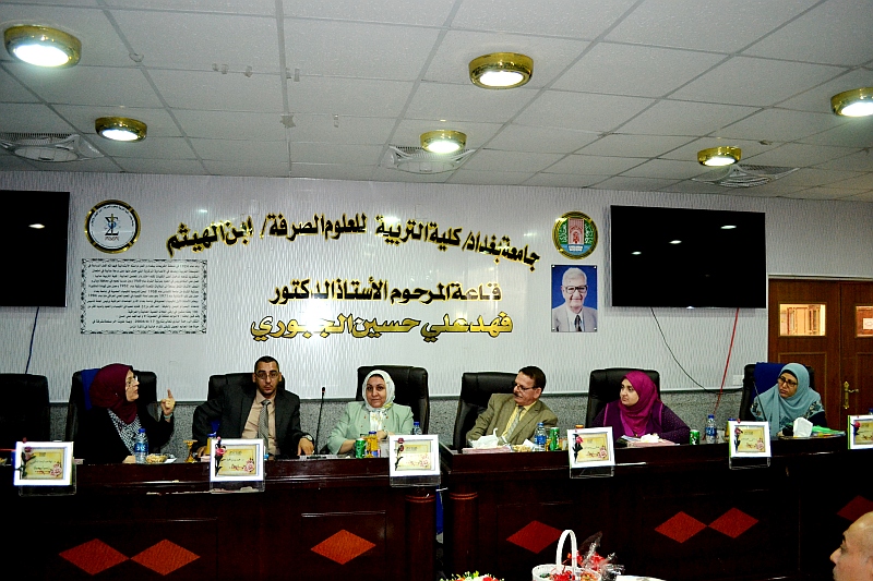
ونوقشت الاطروحة على قاعة المرحوم أ. د. فهد علي حسين من قبل اعضاء لجنة المناقشة المدرجة اسمائهم في ما يلي :

  • أ.د. عدنان وحيد البديري رئيسا.

  • أ.م.د. وجدان بشير عبد عضوا.

  • أ.م.د. محمد وسام المحنة عضوا.

  • أ.م.د. صدامة سعيد فرج عضوا.

  • أ.م.د. اماني محمد جاسم عضوا.

  • أ.د. نهلة عبد الرضا البكري عضوا ومشرفا.

وتهدف الدراسة الى التعرف على :

  • التركيب النسجي للكبد في الدجاج المحلي البالغ.

  • متابعة المراحل المتسلسلة للتكوين الجنيني للكبد في جنين الدجاج المحلي منذ مرحلة ظهوره الأولى وحتى تكونه بالحالة التي يشبه بها البالغ، وتشمل مرحلة قبل الفقس، ومرحلة الفقس ومرحلة بعد الفقس.

  • التعرف على مرحلة ظهور الحموض الأمينية والكلايكوجين والحموض الدهنية في أكباد أجنة الدجاج المحلي والمتمثلة بالاعمار (7، 11، 14، 19) يوماً حضانة ومتابعة وجودهم وفي اليوم (14) بعد الفقس وفي البالغ.

  • ومن خلال مراجعة المصادر المختلفة اتضح عدم توفر دراسات جنينية حول التكوين الجنيني للكبد في الدجاج المحلي Gallus gallus domesticus، ولا حول ظهور الحموض الامينية والكلايكوجين والحموض الدهنية خلال مراحل تكوين الكبد. ونظراً لأهمية هذا النوع من الطيور في الجوانب الاقتصادية ارتأينا اجراء هذه الدراسة للتعرف على التركيب التشريحي والنسجي والتكوين الجنيني للكبد من خلال متابعة المراحل المتسلسلة لتكوينه ومعرفة الاعمار التي تظهر بها الحموض الامينية والكلايكوجين والحموض الدهنية ومتابعة تراكيزهم خلال المدة قبل الفقس وبعده وفي البالغ.

هدفت الدراسة الحالية التعرف على الوصف الشكليائي والتركيب النسجي للكبد وكيس الصفراء في طائر الدجاج المحلي البالغ Gallus gallus domesticus (Linnaeus, 1758)، ودراسة التكوين الجنيني لهما منذ الإشارة الأولى لظهورهما، حصل على أجنة تراوحت أعمارها ما بين (48) ساعة حضانة إلى مرحلة الفقس (21) يوماً، فضلاً عن صيصان بأعمار (1، 7، 14، 21، 28) يوماً كذلك على البالغ أيضاً، وأجريت عليها خطوات التحميل الكامل وتحضير الشرائح النسجية بطريقة شمع البارافين، وقد استعملت خمسة أنواع من الملونات والتي شملت (الهيماتوكسلين هارس-ايوسين، شيف حمض البريوديك PAS، توليدين بلو، فان جيزن وملون البورق القرمزي)، واستعمل المجهر الالكتروني الماسح والنافذ. وحُللت الحموض الامينية والكلايكوجين والحموض الدهنية لأكباد أجنة (7، 11، 14، 19) يوماً حضانة، وصيصان عمر (14) يوماً وكذلك البالغ من الدجاج المحلي.

يتكون الكبد شكليائياً من فصين، الأيمن اهليليجي الشكل وأكبر من الفص الأيسر وذو لون احمر داكن أو بني محمر-بني مائل للاصفرار، ويشغل أغلب أجزاء تجاويف الجسم البطنية الأمامية والوسطى، أما كيس الصفراء فظهر بشكل تركيب كيسي كمثري إلى مغزلي الشكل تقريباً، ذي لون أخضر غامق يقع على السطح الاحشائي للفص الأيمن.

يحاط الكبد نسجياً بمحفظة كليسون المكونة من طبقة حرشفية رقيقة ونسيج ضام كثيف غير منتظم تحتها، تمتد من المحفظة حواجز رقيقة غير عميقة لا تميز الكبد إلى فصيصات، يتكون النسيج الحشوي الكبدي من خلايا كبدية مضلعة الشكل ذات نواة واحدة أو نواتين، تمتلك (1-2) نوية، وأن قمم (3-5) خلايا كبدية هرمية الشكل تحيط بالاقنية الصفراوية، ترتبت الخلايا الكبدية بشكل حبال كبدية شعاعياً حول الوريد المركزي، تحصر بينها فسح الجيبانيات المبطنة بالخلايا البطانية، تحوي تجاويفها على خلايا كبفر وخلايا الدم الحمر ذات نواة، تكون الباحة البابية مدعمة بنسيج ضام ومكونة من فرع للوريد البابي الكبدي، (1-2) فرع للشريان الكبدي و(1-3) فروع لقناة الصفراء وفروع صغيرة من الوعاء اللمفاوي، وجدت الياف شبكية بين الحبال الكبدية، وتجمعات لمفية منتشرة ضمن النسيج الحشوي الكبدي.

يتكون كيس الصفراء نسجياً من ثلاث طبقات، وهي المخاطية ذات طيات والتي تكون في بعض المناطق طيات أولية، ثانوية واخرى ثلاثية، بطانتها الظهارية مكونة من نسيج ظهاري عمودي بسيط ذي حافة مخططة يستند الى صفيحة اصيلة مكونة من نسيج ضام مفكك ذي تجمعات قليلة من الخلايا اللمفية، والطبقة العضلية مؤلفة من ألياف عضلية ملساء مرتبة دائرياً، ثم الطبقة المصلية أو البرانية المؤلفة من نسيج ضام مفكك.

اما جنينياً فأن التكوين الجنيني للكبد في جنين طائر الدجاج المحلي يبدأ عند عمر (48) ساعة حضانة بشكل اندلاق خارجي من الجانب البطني للأديم الباطن للمعي الامامي مكوناً الردب الكبدي الذي يمثل بداءة الكبد ويتألف من نسيج ظهاري عمودي مطبق كاذب حاوٍ على خلايا كأسية وحافة مخططة، يتمايز خلال (48) ساعة حضانة ليكون البرعم الكبدي الذي تتمايز نهايته القاصية إلى ردب كبدي ثانوي ظهري أو أمامي وردب كبدي ثانوي بطني أو خلفي الذي تظهر من نهايته القاصية براعم صغيرة تمتد خلال نسيج اللحمة المتوسطة للحاجز المستعرض ومؤلفة من نسيج ظهاري عمودي بسيط وتتكون براعم صغيرة اخرى من الردب الظهري أو الامامي في جنين عمر (72) ساعة حضانة، وضوح الأسناخ الكبدية وتمايز خلايا الارومة الكبدية ذات الأشكال المختلفة وتصبح بطانة الجيبانيات الكبدية مستمرة، وفي جنين عمر (96) ساعة حضانة يتكون الفص الأيمن للكبد الجنيني وتمايز خلايا الارومة الكبدية الفاتحة والداكنة وظهور القنيات الصفراوية محاطة بــ (8-10) خلايا ارومة كبدية هرمية الشكل، يزداد حجم الفص الأيمن وتكوين الفص الأيسر للكبد الجنيني في جنين عمر (5) ايام حضانة، وتمايزه إلى بدائتين وسطية وجانبية في جنين (6) ايام حضانة وتحول خلايا الارومة الكبدية إلى خلايا كبدية مضلعة الشكل بظهور حبيبات ارجوانية في سايتوبلازم الخلايا الكبدية تمثل الكلايكوجين، أما في جنين (7) ايام حضانة تمايزت الحبال الكبدية بسمك خليتين وظهور القطيرات الدهنية بشكل فجوات صغيرة بيضاء اللون في سايتوبلازمها، وتحول معظم خلايا الظهارة المتوسطة المكعبة الشكل إلى خلايا حرشفية مكونة مع النسيج الضام محفظة كليسون في جنين عمر (8) ايام حضانة وظهور نسيج ضام حول الوريد البابي الكبدي في جنين عمر (10) أيام حضانة، يظهر ضمنه فرع قناة الصفراء في جنين عمر (11) يوماً حضانة فضلاً عن ظهور افراز مادة الصفراء داخل القنية الصفراوية المحاطة بــ (4-8) خلايا كبدية، وظهور فروع من الشريان الكبدي ضمن النسيج الضام في جنين عمر (12) يوماً حضانة، وبذلك تتمايز الباحة البابية في جنين عمر (13) يوماً حضانة، وفي جنين عمر (15) يوماً حضانة تظهر فروع صغيرة للوعاء اللمفاوي وكذلك ظهور خلايا الدم الحبيبية بشكل مجاميع وبمراحل مختلفة من النضج ضمن النسيج الضام المحيط بالباحة البابية، ظهور النسيج الحشوي الكبدي في جنين عمر (17) يوماً حضانة بشكل مصمت مع زيادة الباحات البابية المتكونة في الاجنة بأعمار (19) و(21) يوماً حضانة، مع وضوح إفراز الصفراء في تجويف القنية الصفراوية التي تحاط بـ(3-5) من الخلايا الكبدية، يتميز كبد صوص عمر (7) يوماً بظهور تجمعات لمفية بأحجام مختلفة صغيرة ومتوسطة واخرى كبيرة، وفي صوص عمر (14) و(21) يوماً انتشار التجمعات اللمفية في نسيج الكبد، وفي صوص عمر (28) يوماً اصبح نسيج الكبد يشبه مثيله في البالغ.

أظهرت نتائج الدراسة الجنينية ان التكوين الجنيني لكيس الصفراء يظهر كبداءة في جنين عمر (96) ساعة حضانة على شكل تثخن ظهاري من الأديم الباطن للردب الكبدي مؤلف من خلايا ظهارية عمودية متجمعة مع بعضها، وفي جنين عمر (5) ايام حضانة يظهر كيس الصفراء ملاصقاً مع الجزء المحيطي للفص الأيمن للكبد الجنيني، وذو تجويف ضيق مبطن بنسيج ظهاري عمودي مطبق كاذب ذو حافة مخططة ومحاط بطبقة رقيقة من خلايا اللحمة المتوسطة غير المتمايزة، يصبح موقع الكيس جانبياً ضمن الجزء الجانبي الاحشائي للفص الأيمن في جنين عمر (6) أيام حضانة، وفي جنين عمر (7) ايام حضانة يصبح تجويف كيس الصفراء أوسع ومحاطاً بخلايا اللحمة المتوسطة غير المتمايزة، وفي جنين عمر (9) أيام حضانة ظهرت افرازات الصفراء في تجويف كيس الصفراء وتمايز القليل من خلايا اللحمة المتوسطة إلى ألياف عضلية ملساء ضعيفة التكوين، وفي جنين عمر (10) ايام حضانة اصبح كيس الصفراء كتركيب صغير أسود ضمن السطح الاحشائي للفص الأيمن وبدء تمايز الصفيحة الاصيلة وتمايز الطبقة العضلية على شكل حزم ضعيفة متقطعة مرتبة بصورة دائرية. وكذلك تمايز الطبقة المصلية المحاطة بطبقة من الظهارة المتوسطة، وفي جنين عمر (12) يوماً حضانة ترتبت الألياف العضلية الملساء باتجاهات مختلفة طولية، عرضية ومائلة على شكل حزم متقطعة مرتبة دائرياً، واصبحت البطانة الظهارية للطبقة المخاطية مؤلفة من نسيج عمودي بسيط ذي حافة مخططة في جنين عمر (13) يوماً حضانة مع ظهور طيات قليلة في تجويف الكيس وترتيب ألياف الطبقة العضلية بشكل حزم مستمرة صغيرة دائرياً وتمايز نسيج الطبقة المصلية إلى نسيج ضام مفكك حاوٍ على بعض مقاطع لأوعية دموية صغيرة، وفي جنين عمر (15) يوماً حضانة تمايز الطيات الى أولية وثانوية واخرى ثلاثية في بعض أجزاء جدار الكيس، مع ظهور مقاطع لأوعية دموية صغيرة والقليل من الخلايا اللمفية ضمن طبقة الصفيحة الاصيلة في جنين عمر (18) يوماً حضانة وتمايز طبقات جدار كيس الصفراء الاخرى، واصبح في جنين عمر (21) يوماً حضانة يشبه كيس الصفراء للدجاج البالغ.

شملت الدراسة الحالية أيضاً تحليل الحموض الامينية لأكباد أجنة طائر الدجاج المحلي بأعمار (7، 11، 14، 19) يوماً حضانة وصوص عمر (14) يوماً والبالغ، ووجد (18) حمضاً امينياً هي الاسبارتيك، الكلوتاميك، السيرين، الارجنين، البرولين، الكلايسين، التايروسين، الميثونين، الايزوليوسين، الليوسين، الفنيل النين، الاسبارجين، الالنين، الفالين، الهستدين، اللايسين، السيستين والثريوثنين وكان أعلى تركيز للحموض الامينية في البالغ، وتم قياس تراكيز الكلايكوجين في أكباد أجنة الدجاج المحلي بأعمار (7، 11، 14، 19) يوماً حضانة وصوص عمر (14) يوماً والبالغ، وكان أعلى تركيز له في جنين عمر (19) يوماً حضانة، كذلك تم تحليل الحموض الدهنية في أكباد الأعمار نفسها المذكورة آنفاً ووجدت (5) حموض دهنية وهي حمض البالمتيك، الستياريك، الاوليك، حمض اللينولييك وحمض الفالينولينيك وكان أعلى تركيز للحموض الدهنية في كبد الطائر البالغ.

التوصيات:

  • أفرزت نتائج الدراسة الحالية جملة من التوصيات وكما يأتي:

  • دراسة جنينية مقارنة لتكوين الكبد وكيس الصفراء في فقريات عراقية اخرى.

  • دراسة للتعرف على تأثير بعض المبيدات والهرمونات والعقاقير الطبية البيطرية في التكوين الجنيني للكبد وكيس الصفراء في طائر الدجاج المحلي وفي طيور عراقية اخرى.

  • دراسة حول دور الخلايا الجذعية في عملية تجدد الكبد.

  • دراسة على المستوى الجزيئي لتحديد الجينات المسؤولة عن التكوين الجنيني للكبد وكيس الصفراء في الطيور العراقية وعن التحفيز والتثبيط خلال مراحل التكوين.

  • دراسة التركيب الدقيق للكبد باستعمال المجهر الالكتروني النافذ في فقريات عراقية اخرى.

  • دراسة التركيب الدقيق لكيس الصفراء باستعمال المجهر الالكتروني النافذ لفقريات عراقية اخرى.

  • دراسة بالمجهر الالكتروني الماسح والنافذ لكيس الصفراء في الدجاج المحلي البالغ.

  • دراسة مقارنة بين محتوى الكبد من الحموض الامينية بين طائر الدجاج المحلي وأنواع اخرى من الطيور العراقية وبين الفقريات المختلفة.

  • دراسة تحليل الكلايكوجين في الكبد خلال المراحل الجنينية في أنواع من الطيور العراقية الاخرى.

  • دراسة مقارنة بين محتوى الكبد من الحموض الدهنية بين طائر الدجاج المحلي وأنواع من الطيور العراقية الاخرى.


Department of Biology discusses PhD. Dissertation on   Gallus gallusdomesticu(1758 Linnaeus)by Dalia Hassan Dhahir Ali supervised by prof.Dr.Nahla AbdulRidha Al-Bakri The dissertation was discussed at the hall the late prof.Dr.Fahad Ali Hussein

Summary

The current study aimed to investigate the morphological description and histological structure of the liver and gallbladder in the adult local chicken bird Gallus gallusdomesticus (Linnaeus, 1758), and study the embryonic development of them since the first sign of their appearance. The embryos ranged between (48) hours incubation to the hatching stage (21) days were obtained and the young chicken ages (1, 7, 14, 21, 28) days as well as adult.

It has taken steps of whole amount and preparation of the histological section in the paraffin wax method, and has used five types of stains, which included (Harris Hematoxylin-Eosin, Periodic acid Schiff PAS, Toluidine Bleu, Van Gieson and Borax carmine), and used the scanning and transmission electron microscopes. Amino acids, glycogen and fatty acids of liver embryos at (7, 11, 14, 19) days incubation, young chicken age (14) days as well as the adult of the local chicken were analyzed

The liver morphologically is composed of two lobes, the right elliptic, larger than left lobe, dark red or reddish brown or brown yellowish. It occupies most of the parts of the anterior and middle abdominal cavities. While for the gallbladder, it appeared in the form of cystic pear to spindle almost the shape, dark green and located on the visceral surface of the right lob.

Histologically the liver is surrounded by Glisson capsule, which consists of a thin squamous layer and an irregular dense connective tissue beneath it, stretching from the thin and non-deep septum that do not discriminate the liver to the lobes. The hepatic parenchymal tissue consists of polygonal hepatocytes with one or two nuclei, possessing (1-2) nucleolus,and that the peaks (3-5) of the pyramidal hepatic cells surrounding the bile canaliculi. The hepatocytes cells were arranged in the form of hepatic cords radially around the central vein, confined by the sinusoids spaces that lined endothelial cells and their cavities contained of Kupffer cells and red blood cells with nuclei, the portal area supported by connective tissue and consisted of a branch of the hepatic portal vein, (1-2) branch of the hepatic artery, (1-3) branches of the bile duct and small branches of the lymphatic vessel. Reticular fibers were found between the hepatic cords, and lymphatic aggregations spread within the hepatic parenchymal tissue.

The gallbladder consisted histologically of three layers, which were mucosa with folds that in some areas were primary, secondary and tertiary folds. Epithelial lining of mucosa layer consisting of a simple columnar epithelial tissue with striated border based on lamina proprea consisting of a loose connective tissue with a few aggregations of lymph cells. The muscularis layer were composed of smooth muscle fibers that ranged circularly, then the serosa or adventitia layer consisting of a loose connective tissue

In embryo, the liver development in the local chicken bird begins at the age of (48) hours incubation at an external envagination of the ventral side of the fore gut endoderm, consisting of the hepatic diverticulum, which represents the liver primordium and consists of pseudo-stratified columnar epithelial tissue containing a goblet cells and a striated border. During (48) hours incubation, the hepatic diverticulum was differentiation into hepatic bud, which was differentiated by its distal end into dorsal secondary hepatic diverticulum or anterior and ventral secondary hepatic diverticulum or posterior that appear from the distal end a small buds stretching through the mesenchymal tissue of the transverse septum and composed of simple columnar epithelial tissue. The other small buds composed of dorsal or anterior diverticulum in embryo age (72) hours incubation. In this age, the clarity of hepatic acini and the differentiation of hepatoblast of different forms and the lining of the hepatic sinusoids were continuous. In embryo aged (96) hours incubation the right lobe of the embryonic liver, the differentiation of light and dark hepatoblasts and the appearance of bile canaliculi were surrounded by (8-10) pyramidal hepatoblasts, the right lobe was increased in size and formation of the left lobe of the embryonic liver in embryo age (5) days incubation. The differentiation of left lobes into lateral and medium primordium in embryo (6) day incubation while hepatoblasts were converted into polygonal hepatocytes with the appearance of purple granules in the cytoplasm of the hepatocytes representing the glycogen. Whereas in embryo (7) days incubation, the hepatic cords were differentiated by the thickness of two cells and the appearance of lipids droplets in small white-coloured vacuoles in their cytoplasm. Most of the cubic-shaped mesothelium cells were transformed into squamous cells which formed with the connective tissue Glisson capsule in embryo age (8) days incubation. The connective tissue appearance around hepatic portal vein in embryo age (10) day incubation. The branch of bile duct appeared in embryo age (11) days incubation as well as the appearance of bile secretion inside the bile canaliculi enclosed in (4-8) hepatocytes. The appearance of hepatic artery branches within the connective tissue in embryo age (12) days incubation, thus the portal area in embryo age (13) day incubation. The embryo age (15) day showing small branches of lymphatic vessel as well as the appearance of granular blood cells in the form of groups and at different stages of maturity within the connective tissue surrounding the portal area. The appearance of hepatic parenchymal tissue in embryo age (17) day incubation as compact form with the increase of the portal area in the embryos ages (19) and (21) days incubation, with the clarity of bile secretion in the cavity of bile canaliculi, which is surrounded by (3-5) of hepatocytes. The liver of young chicken age (7) days characterized by the appearance of lymphatic aggregations in different sizes small, medium and large, and in the young chicken age (14) and (21) days showing the propagation of lymphatic aggregations in the liver tissue, and young chicken age (28) day the liver tissue is similar to that of the adult.

The results of the embryonic study showed that the embryo development of the gallbladder appears as a primordium in the embryo age (96) hours incubation in the form of epithelial thickness in the liver diverticulum endoderm composed of columnar epithelial cells accumulated together.

In embryo (5) days incubation, a gallbladder appears adjacent to the peripheral part of the right lobe in the embryonic liver, with a narrow cavity lined with pseudo-stratified columnar epithelial tissue with a striated border and surrounded by a thin layer of undifferentiated mesenchymal cells.

The gallbladder location becomes laterally within the lateral visceral parts of the right lobes of embryo age (6) days incubation. In embryo age (7) days incubation the cavity of gallbladder becomes wider and surrounded by undifferentiated mesenchymal cells. While in embryo age (9) day incubation the bile secretions appeared in the cavity of gallbladder and differentiation a few of mesenchymal cells into poorly formed smooth muscle fibers. In embryo age (10) days incubation the gallbladder became as a small black structure within the visceral surface of the right lobe, starting to differentiate the lamina proprea and differentiate the muscle layer in the form of intermittent thin bundles arranged in a circular shape.As well as the differentiation of the serosa layer surrounded by a layer of mesothelium, and in the embryo of the age (12) days incubation smooth muscle fibers were arranged in different longitudinal directions, transverse and oblique in the form of circular bundles and the epithelial lining of the mucosa layer was composed of a simple columnar tissue with striated border. As well as in the embryo age (13) days incubation the few folds in the gallbladder cavity were appeared, the arrangement of the muscle layer fibers in a continuous small circular bundles and the differentiation of the serosa layer tissue into loose connective tissue contained of some sections of small blood vessels. In embryo (15) days incubation the folds differentiated into primary, secondary and tertiary in some parts of the gallbladder wall, with the appearance of small blood vessel sections and a few lymphocytes within the lamina proprea in embryo age (18) days incubation and differentiation of the rest layer of gallbladder wall. The embryo age (21) days  incubation became like a gallbladder of adult chicken.

The current study also included the analysis of the amino acids in the embryo of a local chicken bird at ages (7, 11, 14, 19) days incubation, young chicken age (14) day and adult, and found (18) amino acid were; Aspartic (Asp), Glutamic (Glu), Serine (Ser), Arginine (Arg), Proline (Pro), Glycine (Gly), Tyrosine (Tyr), Methionine (Met), Isoleucine (Ile), Leucine (Leu), phenyl alanine (Phe), Asparagine (Asn), Alanine (Ala), Valine (Val), Histidine (His), Lysine (Lys),Cysteine (Cys) and Threonine (Thr) and the highest concentration of the amino acid in the adult. The measurement of glycogen in the liver of local chicken embryos at ages (7, 11, 14, 19) day incubation, young chicken age (14) day and adult was carried out and the highest concentration in embryo age (19) days incubation. In addition, the fatty acids in the same ages mentioned above was analyzed and found (5) fatty acids, namely Palmitic, Stearic, Oleic, Linoleic and α-Linolenic acids and the highest concentration of fatty acids in the adult bird liver.

IhcoeduAuthor posts

Avatar for ihcoedu

كلية التربية للعلوم الصرفة (ابن الهيثم) College of Education for Pure Science (Ibn Al-Haitham)

Comments are disabled.