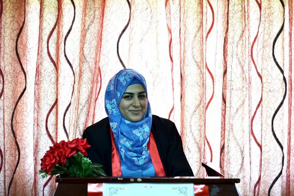
مناقشة رسالة طالبة الماجستير زينب خضير طه من قسم علوم الحياة
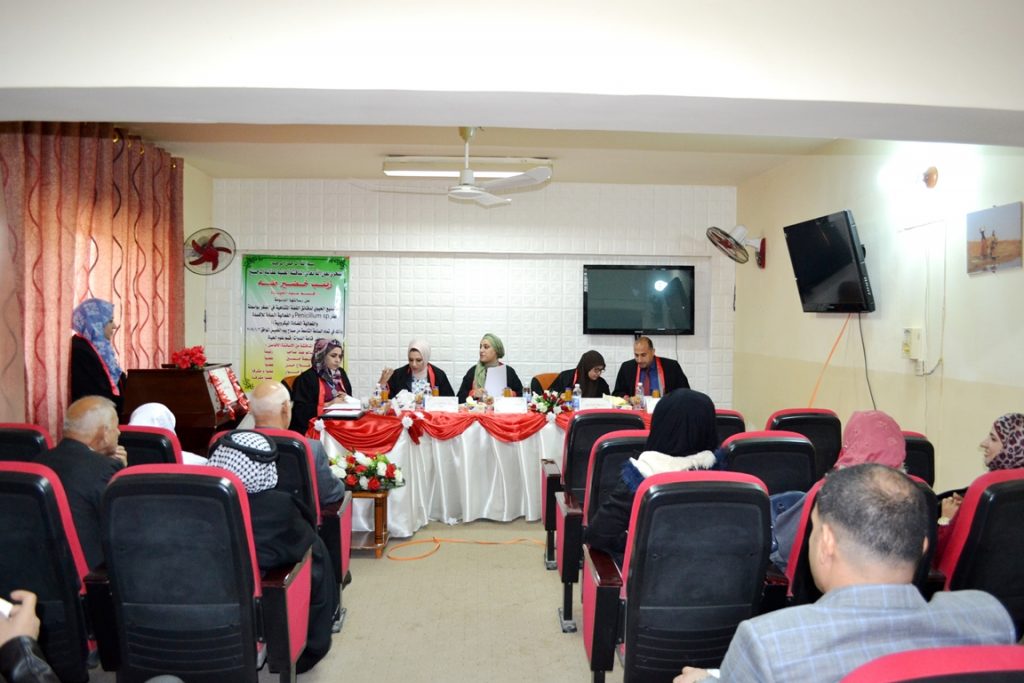
جرت يوم الخميس الموافق 3 / 1/ 2019 على قاعة الدراسات العليا في قسم علوم الحياة المناقشة العلنية لرسالة طالبة الماجستير زينب خضير طه الموسومة :
التصنيع الحيوي لدقائق الفضة المتناهية في الصغر بواسطة فطر Penicillium sp. والفعالية المضادة للاكسدة والفعالية المضادة الميكروبية
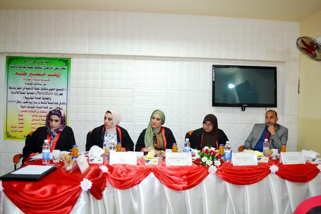
حيث تألفت لجنة المناقشة من التدريسيين الأفاضل :
أ.م.د . هنادي سالم عبد صاحب رئيسا
أ.م.د . نهايةنحس نعمة عضوا
م.د .فادية فلاح حسن عضوا
ا.م.د.سمية نعيمة حوار عضوا ومشرفا
تم في هذه الدراسة تم عزل وتشخيص فطر P.italicum من الليمون الحامض العراقي الذي جمع من الاسواق المحلية في مدينة بغداد ,اذ تم تشخيصه مظهريا بالاعتماد على لون المستعمرة وقوامها وقطرها والتي نميت على ستة انواع من الاوساط الزرعية ( وسط آكار البطاطا والدكستروزPDA , وسط آكار خلاصة الشعير , وسط آكارزابك دوكس , وسط اكار النتريت والسكروز, وسط Czapek Yeast Autolysate (CYA) Agar , آكار سكروز خلاصة الخميرة ) وكذلك مجهريا باستعمال المجهر الضوئي بقوة تكبير 40X وتحديد شكل الكونيدات وشكل الخيوط الفطرية وبمقارنة نمو الفطر في الاوساط الزرعية لوحظ انه هناك فرق معنوي بينها وان افضل الاوساط الزرعية لنمو الفطر هي ( وسط آكار البطاطا والدكستروزPDA , وسط آكار خلاصة الشعير).
بعد تشخيص فطر P.italicum تم اختبارفعاليته في التصنيع الحيوي لجسيمات الفضة النانوية وذلك بمزج الكتلة الحيوية للفطر مع محلول نترات الفضة وملاحظة التغير اللوني الذي يطرأ عليه بعد 96 ساعة ومقارنته مع محاليل السيطرة ,وكانت النتيجة ان المحلول تغير من اللون الاصفر الى اللون البني الداكن كدليل اولي على انتاج جسيمات فضة نانوية بعدها تم توصيف جسيمات الفضة النانوية عن طريق اجراء مجموعة من الفحوصات وهي فحص مطياف الأشعة فوق البنفسجية ( UV / vis spectrophotometer ) اذ ظهرت اعلى قمة لطيف الاشعة فوق البنفسجية عند 415 نانومترا اما فحص مطياف الأشعة تحت الحمراءFourier Transform Infrared Measurement( FTIR) فقد بين وجود امايدات اولية وثانوية في المحلول كدليل على افراز انزيمات وبروتينات من قبل الفطر ساهمت في اختزال ايون الفضة . وعند قياس حيود الأشعة السينية XRD X-Ray Diffraction)) كان التركيب البلوري مكعبي (Cubic) و المستويات البلورية المتكونة هي (111) (200) (202) (311) وبالنسبة لفحص مجهر القوة الذريةAFM Atomic force microscope) ) وجد أن معدل الحجم الحبيبي (Avg. Diameter) كان بحدود81 nm . كما واوضحت نتائج فحص المجهر الالكتروني الماسح Scanning electron microscope) SEM ) أن الحجم الحبيبي يتراوح بين 100-32 نانومتروشكل الجسيمة كروي ونلاحظ من خلال نتائج EDS ان ذرات الفضة تتواجد بنسبة % 74.14 من مجموع مكونات العينة السائلة .
تم اختبار الفعالية المضادة للاكسدة لجسيمات الفضة النانوية بطريقة DPPH واختبار الكسح لصبغة الريسازورين واختبار الجذور الهيدروكسيلية واظهراختبار الكسح لمادة DPPH 53.09% , 46.46% , 43.58% عند التراكيز 300, 200, 100 مايكروغرام /مل على التوالي اذ زادت نسبة الكسح بزيادة التراكيز .بينما اظهر اختبار الكسح لصبغة الريسازورين بان اعلى نسبة كسح كانت %53.09 للتركيز 300 مايكروغرام /مل وعند امتصاصية 3.1 نانومتر مقارنة مع حامض الاسكوربيك .اي ان زيادة تركيز محلول جسيمات الفضة النانوية يزيد من نسبة الكسح للجذور الحرة التي تكونها صبغة الريسازورين .واظهر اختبار الجذور الهيدروكسيلية بان تركيز 300مايكروغرام/مل اعلى نسبة للكسح 53.75% عند امتصاصية 0.148 نانومتر مقارنة مع حامض الاسكوربيك اي بزيادة التركيز تزيد نسبة الكسح للجذور الهيدروكسيلية .
-ستم اختبار الفعالية البايولوجية لجسيمات الفضة النانوية اتجاه البكتيريا والفطريات اذ اظهرت النتائج باستخدام التركيز 1000 , 800 , 600 , 400 مايكروغرام/مل بان اعلى تركيز مثبط كان 1000 مايكروغرام/ مل للانواع البكتيرية Escherichia coli , Staphylococcus aureus , Klebsiella pneumonia حيث بلغ 20 , 17 , 19 ملم للانواع الثلاثة على التوالي .اما بالنسبة للفطريات كذلك كان اعلى تركيز مثبط 1000 مايكروغرام /مل للخمائر Candida albicans , Candida tropicalis و Candida glabrata اذ بلغت نسبة التثبيط35 32 , 35.6 , ملم على التوالي .
-سلوحظ تثبيط واضح في نمو المستعمرات التي تكونها بكتيريا Staphylococcus aureus اكثر من المستعمرات التي تكونها بكتيريا Escherichia coli عند معاملة الوسط بمحلول جسيمات الفضة النانوية اذ كانت نسبة التثبيط على نمو مستعمرات بكتريا S.aureus 90% بينما نسبة تثبيط مستعمرات بكتريا E.coli بلغت 64.5%.
-سفي حين لم يظهر تثبيط في اقطار مستعمرات الفطريات Cladosporum sp Fusarium sp و Monilia sp عند معاملة الوسط الذي تنمو عليه بمحلول الفضة النانوية بالتراكيز 200 , 100 , 50 ,25 مايكروغرام /مل ولكن ظهرت تشوهات في المستعمرات تتمثل بزيادة الغزل الفطري وقلة في الخلايا التكاثرية .
-دعند اختبارتكوين الغشاء الحيوي Biofilm لبكتريا E.coli وخميرة C.Tropicalis باستعمال تراكيز مختلفة من جسيمات الفضة النانوية 1000,800,600 مايكروغرام/مل وقد لوحظ تثبيط في تكوين الغشاء الحيوي يزداد بزيادة التراكيز المستعملة لكل من البكتيريا والخميرة اذ كانت نسب التثبيط (% (84%, 68%,38على التوالي لخميرة C.Tropicalis بينما كانت نسب تثبيط بكتريا (18%,15%,5%) E-coli على التوالي.
-ساجري تحليل تكوين المستعمرات لخلايا سرطان الثدي MCF-7 Clonogenicity assay)) لقياس سمية طويلة الامد لجسيمات الفضة النانوية باستعمال تركيزين 20,10 مايكروغرام التي اظهرت نقصانا في عدد خلايا MCF-7 مقارنة بالسيطرة (خلايا سرطان الثدي MCF-7 الغير معاملة بجسيمات الفضة النانوية) ادت المعاملة بتركيز 10 مايكروغرام /مل الى حدوث تثبيط في نمو خلايا سرطان الثدي وصلت الى 51% بينما كانت نسبة التثبيط اعلى عند تركيز 20 مايكروغرام /مل اذ كانت نسبتها 26%)) عند اجراء اختبارحيوية الخلايا باستعمال صبغه البنفسجي البلوري لملاحظة قدرة الخلايا على البقاء بعد 24 ساعة من الحضن.
The MSc. thesis Discussion of student Zainab Khudair Taha from department of Biology
On Thursday,the 3rd of January 2019,at the hall of higher studies , The MSc. thesis Discussion of student Zainab Khudair Taha from department of Biology entitled (Biosynthesis of Silver Nanoparticles by Fungi Penicilliumsp.Antioxident and Antimicrobial)has been discussed.
The discussion committee
Asst.Prof.Dr. Hanadi Salim Abdulsahib Head
Asst.prof.Dr. Nihaea Nihas Neama member
Asst.Dr.Fadia Falah Hassan member
Asst.prof.Dr.Sumaya Naema Hawar supervisor member
Summary
In this study, P.italicumwas isolated and diagnosed from Iraqi citrus lemonthat collected from the local markets in Baghdad. P.italicumisolated were diagnosed morphologically depending on the appearance of the colony, color and diameter; all isolated were cultured on sixculture media including PDAs,Malt extract agar,Czapekdox agar,Nitrite sucrose (no2) Agar, Czapek Yeast Autolysate (CYA) Agar, and Yeast extract sucrose (YES), as well as isolates were diagnosed microscopically using optical microscope with a magnification of 40X and determine the shape of conidiaand fungal filaments and also comparing the growth of P.italicum in the culture media. The results foundsignificantdifferences in the growth of P.italicumon culture media and the best media for its growth was PDA agar and Malt extract agar media.
After the diagnosis of P.italicum, its activity in the biosynthesis of silver nanoparticles was estimated by mixing the fungus biomass with the silver nitrate solution and observing the color change occurring after 96 hours and comparing it with control solutions. The result was changing of solution from yellow to dark brown as first evidence for biosynthesis of the nanoparticle particles, then the characterized of nanoparticles were determined by a series of tests including UV/vis spectrophotometer, where the highest peak of ultraviolet radiation was observed at 415 nm, while using of Fourier Transform Infrared Measurement (FTIR) showed the presence of primary and secondary amides in the solution as evidence for the secretion of enzymes and proteins by P.italicum that contributed to the reduction of silver ion. Moreover, the measuring of X-Ray Diffraction (XRD), the crystal structure was cubic and the crystalline levels formed were (111) (200) (202) (311),while for Atomic Force Microscope (AFM) it was found that the granular size (Avg. diameter) was up to 81 nm; the results of the scanning electron microscopy (SEM) showed that the particle size ranged between 100-32 nmand the shape of particle was circular, and the results of EDS revealed that the silver atoms are present at 74.14% of the total liquid sample components.
The anti-oxidant activity of silver nanoparticles was determined using DPPH and scavenging test for risazurinedye and hydroxyl radicals. The DPPH scavenging test found that the activity was 46.46%, 53.09%s and 43.58% after using concentrations at 300, 200 and 100 μg/ml and this activity increased with highest concentration. While the scavenging results for risazurine was 53.09% for the concentration of 300 μg/ml and the absorption was at 3.1 nm uptake compared to ascorbic acid. The increases in silver nanoparticle concentration solution increase the scavenging rate for free radicals formed by risazurine dye. The concentration 300 μg / ml given the highest of scavenging percentage 53.75% at 0.148 nm compared to ascorbic acid, concluding the high concentration increased the scavenging activity for hydroxyl radicals.
The biological activity of silver nanoparticle particles was tested against bacteria and fungi. The results showed that after using the concentration of 1000, 800, 600, 400 μg/mlthat the inhibitory concentration was 1000 μg/mlagainstEscherichia coli, Staphylococcus aureus andKlebsiella pneumonia, the inhibition zone was 19, 17 and mm for the three species respectively. For fungi, the highest inhibitory concentration was also 1000 μg/ml against Candida albicans, Candida tropicalis and Candida glabrata, and the inhibition zone was 35, 32 and 35.6 mm respectively.
Moreover, the inhibition that detected against Staphylococcus aureus colonies was more than found with Escherichia coli colonies after the treatment with silver nanoparticles, and the percentage of inhibiting growth of colonies was 90% against S.aureus, while it was 64.5% against E.colicolonies .
However, there was no inhibition in the colonies of Cladosporum sp.,Fusarium sp. and Moniliasp that treated with the solution of the silver nanoparticles with concentrations of 200, 100, 50 and 25 micrograms/ml, but malformations was appeared in the colonies of fungi including the increases of mycelium and decreases in the reproductive cells.
The formation biofilm byE. coli and C.Tropicalisexperiment involved using different concentrations of silver nanoparticle particles (1000, 800, 600 μg/ml)and the results observed an inhibition in the formation of the biomembraneand this inhibition was increased with high concentrations against both bacteria and yeast. The inhibition percentage was 84%, 68% and 38% respectively for C.Tropicalis, while the inhibition percentage forE. coliwas (18%, 15%, 5%) respectively.
The Clonogenicity assay for MCF-7 breast cancer cell line was performed to measure the long-term toxicity of silver nano particle using 20 and 10 μg, the results revealed an inhibition in the number of MCF-7 cells compared to control (MCF-7 breast cancer cells not treated with nanoparticles). Furthermore, the treatment with a concentration of 10 μg/ml resulted in inhibiting in the growth of MCF-7 cells up to 51%, while the inhibitory percentage was up to 26%with 20 μg/ml when testing the cells by using crystalline violet dye to observe the ability of cells to survive after 24 hours of incubation.