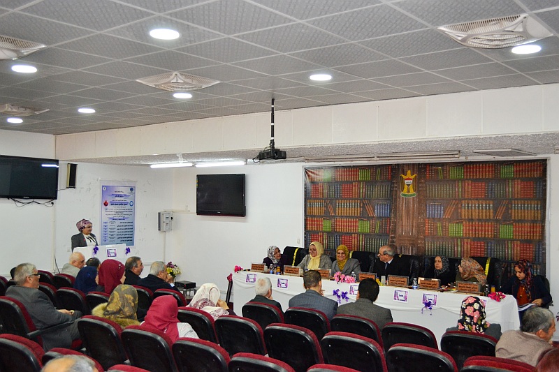
جرت يوم الخميس الموافق 10 / 1/ 2019  وعلى قاعة  المرحوم الاستاذ سالم عبدالحميد  مناقشة اطروحة طالبة الدكتوراه استبرق عزالدين محمود سلمان من قسم علوم الحياة والموسومة :

دراسة نسجية مرضية وكيمونسجية مناعية لمشيمة النساء المصابات بداء السكري


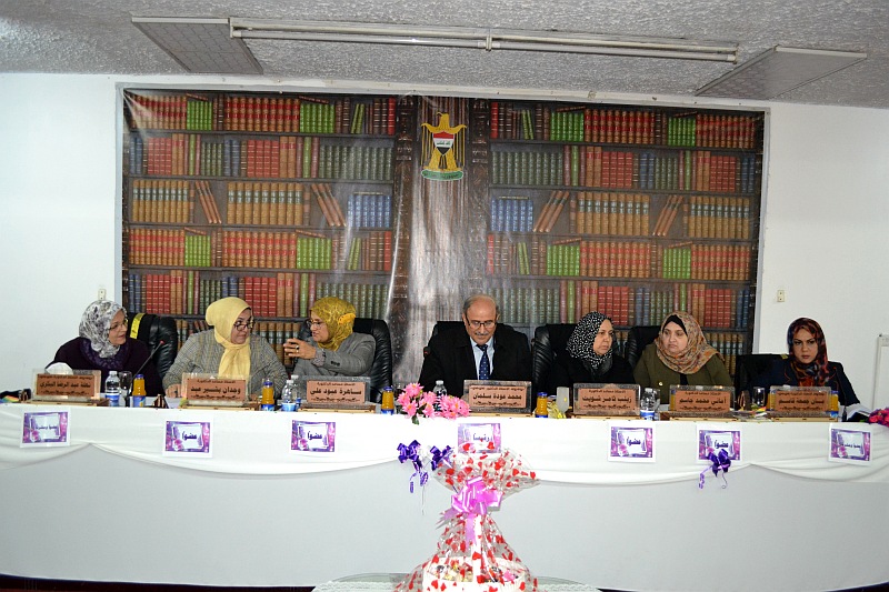
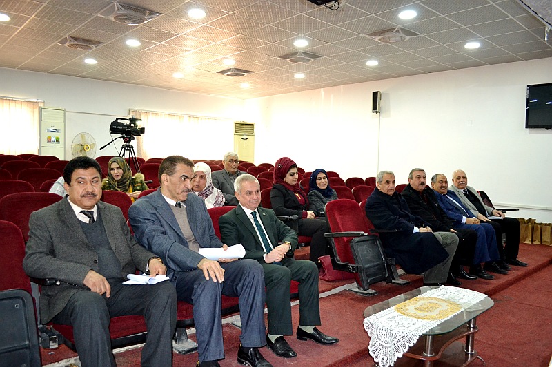
وتألفت لجنة المناقشة من التدريسيين الأفاضل :

ا.د. محمد عودة سلمان                 رئيسا

ا.م.د. ساهرة عبود علي                عضوا

ا.م.د. زينب ثامر شويت                  عضوا

ا.م.د. وجدان بشير عبد                   عضوا

ا.م.د. اماني محمد جاسم              عضوا

ا.د. نهلة عبد الرضا البكري             عضوا ومشرفا

ا.د.  بان جمعة قاسم                     عضوا ومشرفا


  أجريت هذه الدراسة خلال المدة من 2016 الى 2018 في قسم علم الامراض، كلية الطب/جامعة النهرين وقسم علوم الحياة/ كلية التربية للعلوم الصرفة (ابن الهيثم)/ جامعة بغداد. تم الحصول على المشيمات الطرية من قسم النسائية والتوليد في مدينة الامامين الكاظمين (عليهما السلام) الطبية، مستشفى بغداد التعليمي ومستشفى الكرخ للولادة للمدة من 1/12/2016 الى 1/5/2017، وبعد الحصول على الموافقة من وزارة الصحة العراقية. تضمنت الدراسة الحصول على 102 حالة والتي تتضمن 34 حالة مع سكر الحمل و34 حالة مع سكر قبل الحمل و34 حالة مع حمل طبيعي كمجموعة سيطرة. تم في هذه الدراسة المقارنة بين المجاميع الثلاثة على أساس المعلمات الشكليائية، الخصائص النسجية المرضية، القياس النسجي، ترسب الكلايكوجين بمساعدة صبغة حامض البريوديك ﺸﻴف، التعبير عن عامل نمو بطانة الأوعية الدموية-A باعتماد تقنية الكيميائية النسجية المناعية والميزات التركيبية الدقيقة باستعمال المجهر الالكتروني النافذ فضلاً عن تسجيل بعض المعلمات السريرية للام والطفل حديث الولادة.

تظهر نتائج الدراسة زيادة في حدوث سكر الحمل معنوياً (p≤0.05) في المجموعة العمرية 30-39 سنة، مع متوسط عمر 29.82±1.09 سنة. اما عدد مرات الولادة والحمل فقد وجد ان النساء المصابات بالسكري اكثر عرضة لتعدد الولادات والحمل مقارنة مع الحمل لأول مرة. كذلك زيادة حدوث الولادات المبكرة معنوياً (p≤0.05) في مجموعتي سكر الحمل والسكر قبل الحمل بالمقارنة مع مجموعة السيطرة. العمليات القيصرية كانت عالية الحدوث معنوياً (p≤0.001) في مجموعتي السكر (سكر الحمل والسكر قبل الحمل). كان العمر الحملي بتخطيط الصدى غير معنوي (p>0.05) بين المجاميع الثلاثة ولكن موعد اخر دورة حيضية كان معنوياً تحت مستوى احتمالية (p≤0.05).

اظهر حرز ابغار (APGAR score) للوليد الحديث الولادة (بعد 5 دقائق) اختلافاً معنوياً عالياً (p≤0.001) بين المجاميع الثلاثة. كان وزن الوليد حديث الولادة ومؤشر كتلة الجسم (وزن الطفل/وزن الطفل) مختلفاً معنوياً (p≤0.05) ولكن نسبة وزن الجنين/وزن المشيمة كانت غير مختلفة معنوية (p>0.05) بين المجاميع الثلاثة. اما المعلمات الامية فقد كان مستوى متوسط الكلوكوز قبل الولادة غير مرتفع معنوياً (p>0.05) بين المجاميع الثلاثة بينما اخر اختبار خضاب الدم السكري (HbA1c) بين مجموعتي السكري كان عالياً معنوياً (p≤0.001) وسجلت مجموعة السكر قبل الحمل ارتفاع في قيمة هذا الاختبار.

اظهر الوزن والقطر المشيمائي عدم وجود فروق معنوية (p>0.05) بين المجاميع الثلاثة بينما في السماكة المركزية كانت الزيادة معنوية (p≤0.05) وكذلك الحال مع عدد الفلق فقد كانت الزيادة عالية معنوياً (p≤0.001) بين المجاميع الثلاثة. وظهرت علاقة زيادة معنوية عالية (p≤0.001) بين النمط الوعائي الصارم (Magistral) للاوعية الدموية المشيمائية ومجموعتي السكر، بينما النمط المنتشر (Dispersal) لوحظ في النساء الطبيعيات. اما طول وقطر الحبل السري فقد كانت الزيادة المعنوية عالية (p≤0.001) بين المجاميع الثلاثة فضلاً عن انغراس الحبل السري خلال الصفيحة المشيمائية، وسجل لون وعدد الاوردة في الحبل السري زيادة معنوية عالية (p≤0.001) بين مجاميع السيطرة وسكر الحمل والسكر العادي.

سجل عمر الام علاقة مرتفعة معنوياً (p≤0.001) مع الإجهاض او موت الجنين خلال الثلث الأول والثاني من الحمل في مجموعتي السكر معاً، ومع اوزان المشيمة في المجاميع الثلاثة معاً وأنواع الحبل السري في المجاميع الثلاثة معاً وكذلك في مجموعة سكر الحمل لوحدها. فضلاً عن ذلك سجلت نفس العلاقة مرتفعة معنوياً (p≤0.001) بين كل من وزن المشيمة مع وزن الطفل حديث الولادة في المجاميع الثلاثة وطول الحبل السري مع جنس الوليد في المجاميع الثلاثة معاً ومجموعة السكر العادي لوحدها وطول الحبل السري مع كل من وزن الطفل ووزن المشيمة في المجاميع الدراسة الثلاثة معاً، وكذلك مستوى اختبار خضاب الدم السكري مع مجموعتي السكري وطريقة علاج السكري وحالة الوليد مع طريقة علاج الام المصابة بالسكري. بينما لا يوجد علاقة معنوية (p>0.05) بين عمر الام مع جنس الوليد وطول الحبل السري.

الزغابات غير الناضجة، العقد المخلوية، تليف الزغابات، تثخن الغشاء القاعدي للزغابات، الارومة الغاذية الخلوية في الزغابات الانتهائية، تليف خارج الزغابات، وتنخر ليفي، انتشار خلايا هوفباور (Hofbauer) في الزغابات الخلالية غير الناضجة، خلايا الدم الحمراء ذات النواة في الجنين ووذمة الزغابية كانت اكثر حدوثاً في مشيمة مجموعتي السكري بالمقارنة مع المشيمة الطبيعية.

اما القياسات النسجية، سجلت الزغابات الانتهائية واوعيتها الجنينية الدموية في المقطعين المركزي والمحيطي للمشيمات زيادة معنوية عالية (p≤0.001) في قطرها بالنسبة للوليد الذكر مقارنة مع الوليدة الانثى للنساء المصابات بالسكري. فضلاً عن ذلك، اظهر قطر الزغابات الانتهائية في المقطعين المركزي والمحيطي للمشيمات فروق معنوية عالية (p≤0.001) بين المجموعتين الفرعيتين من السكر قبل الحمل (مجموعتي سكر النوع الأول والثاني) ولكن لم تسجل الاوعية الدموية الجنينية في الزغابات الانتهائية اي فروق معنوية (p>0.05).

نتائج الدراسة الكيمونسجية باستعمال صبغة حامض البريوديك ﺸﻴف أظهرت زيادة في ترسب الكلايكوجين بمعنوية عالية (p≤0.001) في مشيمات النساء المصابات بالسكري في كل من داخل الزغابات، حول الاوعية الجنينية والاغشية القاعدية. كذلك سجل المقطعين المركزي والمحيطي للمشيمة اختلافاً في تكرار الصفات الكيمونسجية. 

كان عامل نمو بطانة الأوعية الدموية-A في الخلايا الغاذية والسدى الزغابي والخلايا البطانية للاوعية الدموية الجنينية في المقطع المركزي للمشيمات عالياً معنوياً (p≤0.001) مقارنة مع النساء الطبيعيات في كل من طريقتي التحليل المجهري التقليدي والقياس الرقمي.

وأخيراً كشفت الدراسة التركيبية الدقيقة للمقطع المركزي للمشيمات في مجموعتي السكري انخفاض سمك الغشاء المخلوي-الوعائي وكثافة الزغابات القمية الدقيقة للخلايا الغاذية المخلوية، زيادة سمك الغشاء القاعدي الغذائي، ترسب الكلايكوجين والوذمة.


The PhD. Dissertation Discussion of student (Istbraq Aiz Al-Deen Mahmood)from department of Biology

On Thursday,the 10th  of January 2019,At the hall of the late prof.Dr.Salim Abdulhameed , The PhD. Dissertation Discussion of student (Istbraq Aiz Al-Deen Mahmood)from department of Biology entitled(Histopathological and Immunohistochemical Study of Placenta in Diabetic Women)has been discussed

Abstract

This study was carried out during the period from 2016 to 2018 in the Department of Pathology, College of Medicine/ Al-Nahrain University and Department of Biology, College of Education for Pure Science (Ibn-Haitham)/ University of Baghdad. Fresh placentae were obtained from Department of Obstetrics and Gynaecology in Al-Imamain Al-Kadhimiyain (AS) Medical city, Baghdad Teaching Hospital and Al-Karkh Maternity Hospital for the period 1 December 2016 to 1 May 2017 after obtaining the approval from Iraqi Ministry of Health. A total of 102 cases were included in this study, which includes 34 cases with gestational diabetes, 34 cases with pregestational diabetes and 34 cases with normal pregnancy as a control group. In this study, the placentae of three groups were compared on the basis of morphological parameters, histopathological features, histometrical measurement, glycogen deposition with the assist of periodic acid-Schiff stain, expression of vascular endothelium growth factor-A by using immunohistochemical technique and ultrastructural features by Transmission Electron Microscope as well as registered of some clinical parameters of mothers and neonates.

The results of this study showed an increased incidence of gestational diabetes significantly (p≤0.05) in the age group of 30-39 years, with the mean agewas 29.82±1.09 years. As for parity and gravida, multiparous and gravida were have been found to be more susceptible for diabetes mellitus women in comparison to the primigravida. As well as increased was occurred preterm significantly (p≤0.05) in gestational and pregestational diabetes groups in compression to control group. Caesarean section was a highly significant occurrence(p≤0.001) inthe two diabetic groups. Gestational age at ultrasonography was no significant (p>0.05) among the three groups, but at the last menstrual period was significant at the probability level (p≤0.05).

APGAR score of the neonate (after 5 min) demonstrated a highly significant increase(p≤0.001) among the three groups.Neonate weight and BMI (the weight of a baby/ length of the baby) have been significant increases(p≤0. 05), but fetal/placental weight ratio was a non significant increase (p>0.05) among the three groups.  As for maternal parameters, mean glucose levels before labour was a non significant increase (p>0.05) among the three studied groups, whereas last maternal glycosylated hemoglobin A1c between two diabetic groups showinga highly significant increase (p≤0.001) and the pregestational diabetesgroup recorded evaluation in last maternal glycosylated hemoglobin A1c value.

Placental weight and diameter showed non significant increases (p>0.05) among the three groups, while central thickness has been a significant increase (p≤0.05) so as the number of cotyledons were highly significant increase (p≤0.001) among the three studied groups. Magistralvascular pattern of chorionic blood vessels shown ahighly significant (p≤0.001) relationship with the two diabetic groups, whilst dispersal pattern was seen in normal women. The umbilical cord long and diameter have been highly significant increases(p≤0.001) among the three groups as well as umbilical cordinsertion onto the chorionic plate, its colour and number of veins in umbilical cord recorded highly significant increases (p≤0.001) among control, gestational and pregestational diabetes groups

Maternal age of the studied groups recorded a highly significant (p≤0.001) in relationship to the first and second trimester of miscarriage or stillbirths in the two diabetic groups together, placental weights in the three studied groups together and umbilical cord types in the three studied groups together and gestational diabetes group separately. Additionally, same highly significant (p≤0.001) relationship was recorded between each of placental weight with neonatal weight in the three studied groups, umbilical cord length with the gender of neonate in three studied groups together and pregestational diabetes group separately and umbilical cord length with the weight of each neonate and placenta in the three studied groups together, last maternal glycosylated hemoglobin A1c level with the two diabetes groups and method of its treatment, state of the neonate with the method of treatment in diabetic mother. While no significant (p>0.05) relationship was found between maternal age with neonatal gender and umbilical cord lengths.

Immature villi, syncytial knots, villous fibrosis, basement membrane thickening of villi, cytotrophoblast in terminal villi, extravillous fibrinoid, fibrinoid necrosis, Hofbauer cell population in immature intermediate villi, calcification, chorangiosis, thrombosis and thickening of villi vessels, nucleated fetal red blood cells and villous edema have been more occurrence in placenta of the two diabetic groups in comparison to the normal placenta.

Histometrically, terminal villi and their fetal blood vessels of central and peripheral sections of placentae recorded mostly highly significant increases (p≤0.001) of diameters in male neonates as compared with female neonates of diabetic women. Moreover, the terminal villi diameter in central and peripheral sections of placentae appeared highly significant differences (p≤0.001) between pregestational diabetes type 1 and type 2, but the diameter of fetal blood vessels in this terminal villous did not record any significant difference (p>0.05).

The results of the histochemical study by of periodic acid-Schiff stain showed an increase in glycogen deposition with high significance (p≤0.001) in the placentae of diabetic women within intravillous core, around the fetal vessels and basement membranes. As well as central and peripheral sections of placenta were registered difference in frequency of histochemical features.

Vascular endothelium growth factor-A in the trophoblastic cells, villous stroma and fetal vessel endothelial cells in the central section of placentae in the two diabetic women groups have been significantly increased (p≤0.001) compared to the normal women in both manual microscopic analysis and digital quantification.

Finally, ultra structural study of the central section in the placentae of two diabetic women groups revealed a decreased the thickness of vascular-syncytial membrane and density of syncytiotrophoblast apical microvilli, increased thickness of trophoblastic basement membrane, glycogen deposits and edema.

IhcoeduAuthor posts

Avatar for ihcoedu

كلية التربية للعلوم الصرفة (ابن الهيثم) College of Education for Pure Science (Ibn Al-Haitham)

Comments are disabled.