

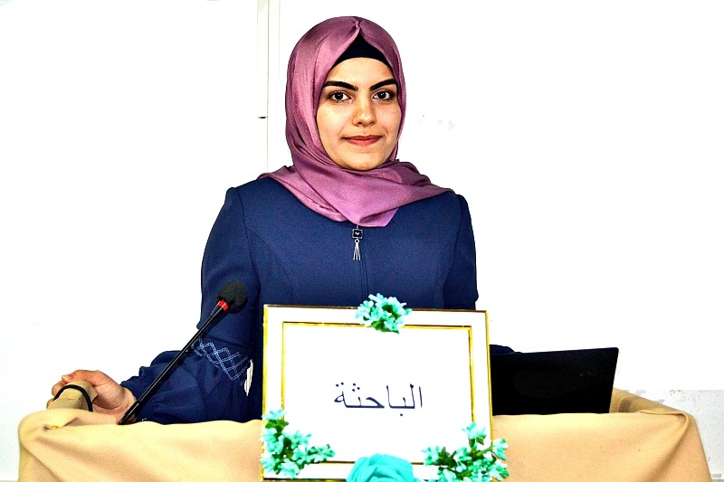
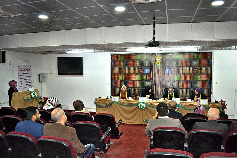
ناقش قسم علوم الحياة في كلية التربية للعلوم الصرفة (ابن الهيثم) رسالة الماجستير الموسومة ” الوصف الشكليائي والتركيب النسجي للرغامي والرئة في الخفاش” للطالبة (حنين إبراهيم الحياني) التي انجزت باشراف التدريسية في القسم (أ.م.د. بيداء حسين مطلك) حيث تمت المناقشة على قاعة المرحوم (أ. سالم عبد الحميد).
وتم أجراء الدراسة على (10) نماذج من الخفافيش عارية العجز كان معدل وزنها بين 45.8 -53 غم وبعد استئصال الرغامي والرئة ثبتت بمحاليل التثبيت واجريت عليها الخطوات المتسلسلة في تحضير الشرائح النسجية وتم استعمال اربعة انواع من الملونات متمثلة بالملون الروتيني الهيماتوكسلين والايوسين وملون شف حامض البريودك ، وماسون ثلاثي الكروم وملون احمر الاليزارين .
و اظهرت نتائج الدراسة الحالية ان الرغامي في الخفاش عاري العجز يبدو كأنبوب غضروفي مرن غير قابل للانطواء , يمتد من نهاية الغضاريف الحلقية للحنجرة في مستوى الفقرة العنقية الثانية عند منطقة ارتباط الغدة الدرقية في السطح البطني للحلقات الغضروفية الثلاثة الاولى, بعد ذلك يتفرع الى فرعين من القصبات الهوائية الاولية تدخل احداها الى الرئة اليمنى والثانية الى الرئة اليسرى . يتألف انبوب الرغامي من سلسلة من الحلقات الغضروفية في الخفاش تظهر غير كاملة وبشكل حرف C والتي تفتح في السطح الخلفي المقابل للمريء ويربط نهايتي الحلقات الغضروفية نسيج ضام مفكك وحزم من الالياف العضلية الملساء والمتمثلة بالعضلة الرغامية.
بلغ عدد الحلقات الغضروفية حوالي 17-15 حلقة غضروفية والتي تم عدها باستعمال ملون المثيلين الازرق ، يقسم الرغامي على ثلاثة اجزاء الجزء العلوي, الجزء الوسطي والجزء السفلي وبلغ معدل قطر الجزء العلوي للرغامي 1190.82±69.51مايكروميتر اذ كان اقل قطرا من الجزء الوسطي والجزء السفلي اذ بلغ معدل قطر الجزء الوسطي 1635.56±101.24 مايكروميتر ، اما معدل قطر الجزء السفلي فهو اكبر من معدل قطر الجزء العلوي والوسطي و بلغ 1681.53±54.40 مايكروميتر. يتألف جدار الرغامي نسجيا من ثلاثة غلالات متمثلة بالغلالة المخاطية , الغلالة تحت المخاطية والغلالة البرانية الحاوية على الحلقات الغضروفية. تتألف الغلالة المخاطية من ظهارة مطبقة عمودية كاذبة مهدبة حاوية على ثلاث انواع من الخلايا وهي الخلايا العمودية، الخلايا القاعدية والخلايا الكأسية والتي تظهر منتشرة بشكل قليل في الجزء الوسطي والجزء السفلي ما بين الخلايا العمدية المهدبة.
وبناء على ما تقدم فان الخفاش عاري العجز يمتلك زوجاً من الرئات الاسفنجية التي تحتل معظم التجويف الصدري وتحتضن القلب وتظهر كلا الرئتين بلون وردي محمر ومغطاة بغشاء الجنب الحشوي , وتبدو هرمية الشكل ذات سطح املس ، وتمتلك كل رئة قمة قحفية حادة ومطوية الى الاسفل في الرئة اليمنى وشبه مستديرة في الرئة اليسرى وتمثل هذه القمة الفص القمي اما القاعدة الذنبية لكل رئة فقد ظهرت مستديرة الشكل ومطوية في الرئة اليمنى وتمثل الفص القاعدي لكلا الرئتين, ظهرت الرئة اليسرى للخفاش عاري العجز موضوع الدراسة الحالية اصغر من الرئة اليمنى وتتألف من ثلاثة فصوص فص قمي وفص وسطي وفص قاعدي ، يقسم الفص الوسطي على فصيصات صغيرة مشطورة من الوسط , اما الرئة اليمنى فهي اكبر من الرئة اليسرى وتتألف من اربعة فصوص فص قمي وفصين وسطيين وفص قاعدي منطوي الى الاعلى. اظهرت نتائج الدراسة الحالية للخفاش عاري العجز ان الرغامي تتفرع على فرعين من القصبات الهوائية الاولية التي تكون مشابهة في تركيبها النسجي للرغامي , اذ تتألف الغلالة المخاطية من نسيج ظهاري مهدب, اما الطبقة تحت المخاطية فهي تحوي على غدد مخاطية وغدد مصلية والغلالة البرانية تميزت بوجود الغضروف الزجاجي . تتفرع القصبة الهوائية الاولية الى قصبات ثانوية وثالثية ويكون الغضروف الزجاجي فيها بشكل صفائح مقطعة غير منتظمة وتبدو الغلالة المخاطية بشكل طيات اصبعية حاوية على خلايا عمودية طويلة والخلايا الكأسية قليلة العدد وتنفصل الغلالة المخاطية عن الغضروف الزجاجي بطبقة من الالياف العضلية الملساء ,اظهرت النتائج ان القصيبات الاولية كانت اكبر قطرا من الاقطار الاخرى جاءت بعدها القصبات الثانوية ثم القصبات الثالثية فالقصيبات والقصيبات التنفسية والتي لم يكن بين اقطارها فرق معنوي بعدها القصيبات النهائية والقنوات السنخية والاكياس السخية واخيراً الاسناخ .
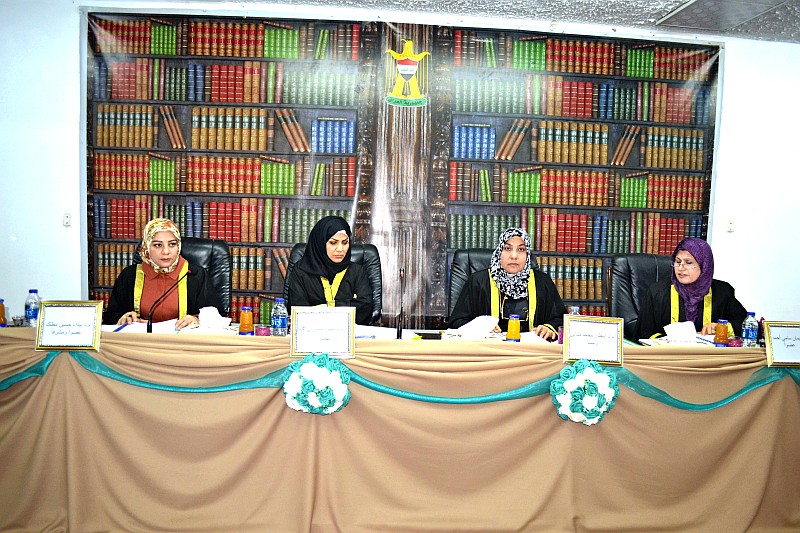
وحصلت الطالبة على تقدير (امتياز) لدى مناقشتة رسالتها من قبل لجنة المناقشة التي تألفت من الذوات :
-
أ.م.د. انتضار محمد مناتي (رئيسا).
-
أ.م.د. سيناء جبوري محمد (عضوا)
-
أ.م.د. ايمان سامي محمد (عضوا).
-
أ.م.د. بيداء حسين مطلك (عضوا و مشرفا)
MSc.thesis on Morphological Description and Histological Structure of the Trachea and Lung in Bat
The department of Biolog held a discussion of MSc.theis of student Hanin Ibrahim Al-Haiani on (Morphological Description and Histological Structure of the Trachea and Lung in Bat) supervised by Asst.prof.Dr.Baida’a Hussein Mutlak
The discussion committee
Asst.prof.Dr.Intdhar MohammedMunati head
Asst.prof.Dr.Sina’a Jaboori Mohammed member
Asst.prof.Dr.Eman Sami Mohammed member
Asst.prof.Dr.Baida’a Hussein Mutlak supervisor member
The Abstract
The current study included the morphological description and the histological structure of the trachea and the lung of thenaked-rumped tomb bat (Taphozousnudiventris) and the results showed the following
This study was conducted on (10) samples of naked-ramped tomb bats whose weight rate was between 45.8-53 gm. After removal of the trachea and the lung, they were fixed with the fixation solutions and performed the series steps in the preparation of the histological sections for the histological study using four types of stain shematoxylin and eosin, special stains (Schiff periodic acid (PAS), Masson´s trachoma, and Alizarin red stain
The results of the current study showed that the trachea in naked-ramped tomb bat was seemed as cartilaginous, flexible and non-foldable tube, extending from the end of the ringed cartilage of the throat at the level of the second cervical vertebra at the thyroid link area of the abdominal surface of the first three cartilage rings. Then it was divided into two branches of the primary bronchi, one entering the right lung and the other to the left lung. The tracheal tube consisted of a series of cartilage rings in the bat that appear in complete and in the form of C-shaped, which open in the posterior surface of the esophagus and connect the ends of the cartilage rings to a loose connective tissue and bundles of smooth muscle fibers represented by the tracheal muscle
The number of cartilage rings were around 15-17 rings, which was counted using the methy lene blue stain and prepared in the laboratory. The trachea was divided into three parts of the upper part, the middle part, the lower or caudal part. The mean diameter of trachea bat was (1190.8 2 ± 69.51 mm). It was less diameter than the middle part and the lower part where the mean diameter of the middle part (1635.56 ± 101.24 mm), while the mean diameter of the lower part was greater than the mean diameter of the upper and middle parts which recorded (1681.53 ± 54.40 mm). This difference was related to the structural synthesis and different sizes of the animals based on the requirements of functional need. The tracheal wall consisted of three tunicae representing of tunic mucosa, tunica sub mucosa and tunica adventitia containing the cartilage rings. Tunica mucosa consisted of a pseudo stratified ciliated columna repithelium with a container on three types of cells, namely columnar, basal and goblet cells. The goblet cells were scattered lightly in the middle and lower parts within the ciliated columnar cells.
The naked-rumped tomb bat hada pair of spongy lungs that occupy most of the thoraciccavity and embrace the heart. The both lungs showing reddishp inkcolor and covered with the visceralpleurisy. As well as it was a smooth-surface pyramid and each lung had an acute cranial apex and it was folded down in the right lung and semi-round in the left lung. This apex represents the apical lobe, and the caudal base of each lung appeared round-shaped, folded in the right lung, and represents the basal lobe of both lungs.
The left lung of the naked-ramped tomb bat in current study was appeared smaller than the right lung and consists of three lobes of apical middle and the basal lobes. The middle lobe, divided into a smalllobular, ripped in the center. The right lung was larger than the left lung and consisted of four lobes, apical, two middle and basal lobes folded into the upper. The results of the current study of the naked-ramped tomb bat had shown that the trachea was branched into two branches of the primary bronchus, which were similar in their histological structure to the trachea. The tunic mucosa consisted of epithelial ciliated tissue containing columnar, basal and goblet cells, while tunica sub mucosa contained of mucous and serosa glands, and the tunica adventitia characterized by the presence of hyaline cartilage.
The primary trachea was branched into secondary and tertiary trach eales where the hyaline cartilage was in the form of irregular sectioning plates and the tunica mucosa appeared as a finger-shape container on long columnar cells and low-number of goblet cells which separated from the hyaline cartilage a layer of smooth muscle fiber.
The results of the current study showed that the primary bronchus were the largest diameter of the rest of the diameters followed by secondary bronchus, bronchioles and respiratory bronchioles, which there was no significant difference between the final bronchioles and the alveolar sac. The previous studies had shown that the bronchioles diameters played important role in airflow to all parts of the lung and the diameter of the bronchioles varies depending on airflow