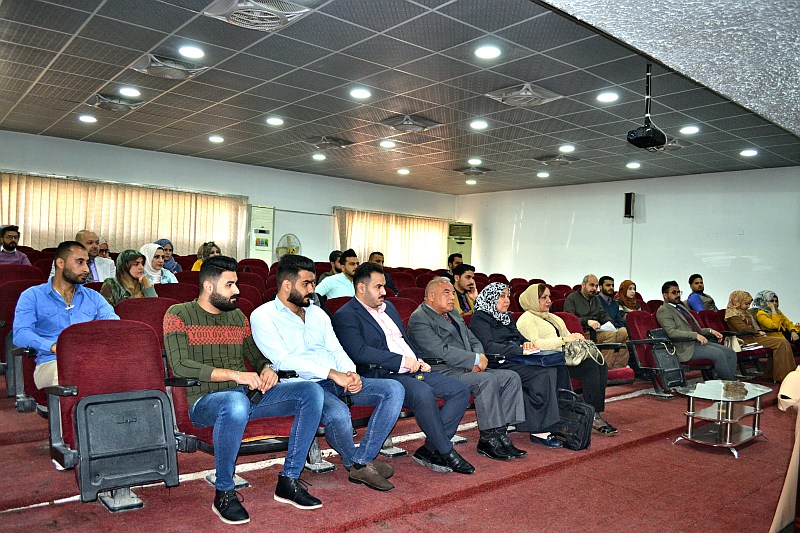
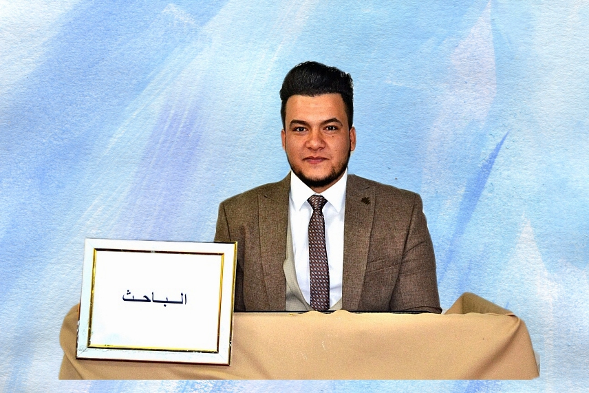
ناقش قسم علوم الحياة في كلية التربية للعلوم الصرفة (ابن الهيثم) بجامعة بغداد رسالة الماجستير الموسومة (دراسة شكليانسجية مقارنة للمصفار في نوعين من الطيور العراقية) للطالب (ياسر سلام كريم ياسين) التي انجزت باشراف التدريسية في القسم (ا.م.د. ايمان سامي احمد) .

تهدف الدراسة الى الـــــــى دراســــــة مصــــــفار الطيـــــــور العراقية وخصوصا الـــــــبرية غير المــــــدروسة والمهددة بالانقراض حيث تم اختيار نوعين من الطيور العراقية  تنتميان الى عائلتين مختلفتين متمثلة بطائر السلوى و اليمام المطوق للتعرف على الوصف الشكليائي والتركيب النسجي للمصفار في هذين النوعين.

واظهرت النتائج ما يأتي :-

اولا / فيما يخص الوصف الشكليائي:

* يكون المصفار في طائري موضوع الدراسة من النوع الرغاموي القصبي يتموضع في الجزء القحفي من التجويف الصدري البطني عند منطقة تفرع الرغامى الى قصبتين ويتخذ موقعاً وسطياً بين القلب والمريء.

* يتألف المصفار من تركيبين , تركيب صلد واخر طري يتمثل الاول بمكونات هيكلية تتضمن الجزء القحفي(الجزء الرغاموي المصفاري) , والجزء الوسطي( الطبلة) و الجزء الذيلي(الجزء القصبي المصفاري) و اخيراً الوتد . فيما يتمثل الثاني بتراكيب غشائية تشتمل, اغشية الطبلة الجانبية , اغشية الطبلة الوسطية, فضلاً عن الرباط بين القصبي الذي يعد مع العضلات المرتبط بالمصفار تراكيب ملحقة للمكونات الرئيسة .

* يتمثل كل من الجزء القحفي و الذيلي لمصفار كلا الطائرين  بأربع حلقات غضروفية فيما تبين ان الجزء الوسطي لطائر السلوى يتمثل بحلقة قحفية مزدوجة وزوج من الحلقات الذيلية في حين يكون لطائر اليمام المطوق حلقة قحفية و اخرى ذيلية .

* تتخذ الطبلة في طائر السلوى شكلاً كالمظلة في حين تظهر في طائر اليمام المطوق اسطوانية الشكل اما الوتد فيظهر اسفينياً في كل من الطائرين.

* يظهر لطائري موضوع الدراسة الحالية زوجان من اغشية الطبلة , يتموضع الزوج الاول على جانبي حلقات الطبلة ومن جهتها الخارجية تدعى بأغشية الطبلة الجانبية فيما يتموضع الزوج الثاني عند الجانب الداخلي لكل فرعي الجزء القصبي المصفاري وتدعى بأغشية الطبلة الوسطية.

* يمتلك مصفار طائر السلوى زوجين من العضلات الخارجية في حين يظهر لطائر اليمام المطوق زوجان من العضلات الخارجية و زوجان من العضلات الداخلية وتحتضن الاخيرة الجزء الوسطي من المصفار لتعطيه شكلاً برميلياً.

* يقع بين فرعي الجزء القصبي المصفاري لطائري موضوع الدراسة الحالية تركيب يدعى بالرباط بين القصبي.

ثانيا / فيما يخص التركيب النسجي:

* اظهرت نتائج الدراسة الحالية ان المصفار في الطائرين يتألف من ثلاث غلالات متمثلة بالغلالة المخاطية المتضمنة البطانة الظهارية , وغلالة الصفيحة الاصيلة- تحت المخاطية و الغلالة البرانية .

* تتألف البطانة الظهارية للجزء الرغاموي المصفاري لطائر السلوى من نسيج عمودي مطبق كاذب مهدب في حين يكون من النوع الظهاري الحرشفي المطبق غير المتقرن في اليمام المطوق اما غلالة الصفيحة الاصيلة- تحت المخاطية فأنها مؤلفة من نسيج ضام مفكك في كلا الطائرين كما تبدو الغلالة البرانية مؤلفة من نسيج ضام مفكك تتخلله اوعية دموية و اعصاب .

* تتألف البطانة الظهارية للجزء القصبي المصفاري في طائري موضوع الدراسة من نسيج عمودي مطبق كاذب مهدب فيما تظهر في غلالة الصفيحة الاصيلة- تحت المخاطية و الغلالة البرانية من نسيج ضام مفكك لكلا الطائرين ايضاً.

* ان البطانة الظهارية لوتد مصفار طائري السلوى و اليمام المطوق تتألف من نسيج عمودي مطبق كاذب مهدب فيما تتألف غلالة الصفيحة الاصيلة- تحت المخاطية من نسيج ضام مفكك في ذكور واناث طائر السلوى الا انه يكون مؤلفاً من نسيج ضام كثيف في الذكور ومفككاً في اناث اليمام المطوق كما تبين ان هيكل الوتد في كلا طائري موضوع الدراسة يكون غضروفياً في الاناث و متعظماً في الذكور.

* تبين ان البطانة الظهارية لكلا غشائي الطبلة الجانبي و الوسطي لطائر السلوى مؤلفة من نسيج ظهاري عمودي مطبق كاذب غير مهدب في حين تكون في اليمام المطوق مؤلفة من نسيج ظهاري حرشفي مطبق فيما تظهر غلالة الصفيحة الاصيلة- تحت المخاطية لغشائي الطبلة في طائري موضوع الدراسة مؤلفة من نسيج ضام كثيف, كما تبين ان حلقات الطبلة تتمثل بغضاريف زجاجية في طائر السلوى فيما يبدو فيها التعظم في ذكور اليمام المطوق فقط .

* تتألف بطانة الشفة الجانبية لطائر السلوى من نسيج ظهاري مطبق مكعب في حين تظهر مؤلفة من نسيج ظهاري حرشفي مطبق في اليمام المطوق , اما بخصوص غلالة الصفيحة الاصيلة- تحت المخاطية فتبين ان النسيج في طائري موضوع الدراسة يتألف من نسيج ضام كثيف.

* تتألف البطانة الظهارية للشفة الوسطية من نسيج ظهاري حرشفي مطبق في طائر السلوى في حين يكون مؤلفاً من نسيج ظهاري عمودي مطبق كاذب في اليمام المطوق اما فيما يخص غلالة الصفيحة الاصيلة- تحت المخاطية فأنها تتألف من نسيج ضام كثيف غير منتظم في طائر السلوى الا انه يكون مؤلفاً من نسيج ضام كثيف منتظم في اليمام المطوق.

* يتباين توزيع او انعدام الخلايا الكأسية والغدد السنخية البسيطة بين النسيج الظهاري لأجزاء المصفار بين طائري موضوع الدراسة الحالية لكنه اتضح بشكل ملفت للنظر انعدام الخلايا الكأسية والغدد السنخية في الشفة الوسطية لطائر السلوى في حين تبين كثافتها في طائر اليمام المطوق .

* كما تباين توزيع الالياف المطاطة والمغراوية المتواجدة في غلالة الصفيحة الاصيلة- تحت المخاطية في اجزاء مصفار كلا الطائرين موضوع الدراسة اذ اوضحت النتائج تركز الالياف المطاطة تحت الظهارة في غشاء الطبلة الوسطي لطائر السلوى في حين تتركز الالياف المطاطة تحت الظهارة في اليمام المطوق ، فيما تبين تواجد الالياف المغراوية بكثافة تحت ظهارة غشاء الشفة الجانبي لكلا الطائرين موضوع الدراسة الا انهما تباينا في الترتيب اذ كان ترتيبهما بالاتجاه البطني – الظهري في طائر السلوى بينما كان ترتيبهما قحفياً- ذيلياً في اليمام المطوق اما غشاء الشفة الوسطي فقد كانت كثافة الالياف المطاطة نحو الخارج وتترتب بالاتجاه القحفي-الذيلي في طائر السلوى في حين كانت كثافة الالياف المغراوية تحت الظهارة و بالاتجاه القحفي – الذيلي في اليمام المطوق.

ثالثا / فيما يخص الدراسة الاحصائية :

* اظهرت نتائج الدراسة الحالية وجود فروق معنوية (0.001>P ) في معدل سمك الغلالات في طائر السلوى لكل من الجزء الرغاموي المصفاري و غشاء الطبلة الجانبي والجزء القصبي المصفاري اذ تكون اكثر سمكاً في الذكور مقارنةً مع الاناث كما تبين وجود فروق معنوية (0.001>P ) في معدل سمك الغلالات  للشفة الجانبية و الوسطية التي تكون في الذكور اعلى مما هي عليه في الاناث و على عكس ذلك تبين وجود فروق معنوية في معدل سمك غشاء الطبلة الوسطي و الوتد اذ تكون اكثر سمكاً في الاناث مقارنةً مع ذكور الطائر نفسة.

* اما طائر اليمام المطوق فقد تبين عدم وجود فروق معنوية بين الذكور و الاناث للجزء الرغاموي المصفاري و القصبي المصفاري و غشاء الطبلة الجانبي في حين وجدت فروق معنوية (0.001>P ) في الشفة الجانبية و غشاء الطبلة الوسطي و الوتد اذ تبين معدل سمك الغلالات في الاناث كانت هي الاعلى مما في الذكور .

* لم تظهر اية فروق معنوية عند مقارنة ذكور طائري الدراسة الحالية و كذلك الحال بين اناثهما في كل من الجزء الرغاموي المصفاري و القصبي المصفاري و غشاء الطبلة الجانبي في حين وجدت فروق معنوية (0.001>P ) بين ذكور الطائرين في غشاء الطبلة الوسطي اذ تكون في ذكور السلوى اسمك مما هي علية في ذكور اليمام المطوق و على العكس من ذلك تكون الشفة الجانبية و الوتد في ذكور اليمام المطوق هي الأسمك.

*تبين وجود فروق معنوية (0.001>P ) بين اناث طائري الدراسة الحالية في كل من الشفة الجانبية و الوسطية و غشاء الطبلة الوسطي اذ تبين ان اناث اليمام المطوق تكون هي الأسمك مقارنةً بإناث السلوى.

افرزت نتائج الدراسة الحالية لمصفار طائري السلوى و اليمام المطوق مجموعة من النقاط تحتاج الى المزيد من الدراسة و التقصي لأجل الوصول الى الحقائق العلمية و بناءً الى ذلك اوصت الدراسة بما يأتي :-

1- اجراء دراسات اخرى على مصفار الطيور العراقية التي لم تنل الاهتمام العلمي الكافي.

2- اجراء دراسة معمقة في ترتيب الالياف المغراوية و المطاطة ضمن الاغشية المهتزة باستعمال ملونات خاصة .

3- قياس ابعاد تجويف و سمك الحلقات الغضروفية لأجزاء المصفار.

4- اجراء دراسة معمقة لمكونات العضلات و عن كيفية ارتباطها بأجزاء المصفار.

5- استعمال ملون ماسون ثلاثي الكروم Masson’s Trichrome للتحري عن الالياف المغراوية Collagen Fibers .

6- استعمال ملون الاليشين الازرق PH(1) Alcian Blue و PH(2.5) للتحري عن الكربوهيدرات القاعدية و الحامضية Sulfated and Acidic Carbohydrates.   

7- تحديد ميزات Glycohistochemistry في اغشية الطبلة الجانبية و الوسطية باستعمال مقاطع مجمدة Frozen Sections للتحري عن انواع اللكتينات Lectins (Carbohydrates- binding Proteins).

8- اجراء دراسات اخرى لمعرفة التجهيز الدموي و العصبي للمصفار .

9-قياس مدى الترددات الصوتية في الطيور وعلاقتها مع التركيب لتتكامل الصورة حول الموضوع.

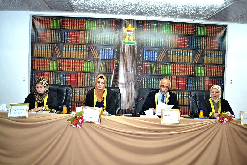
وحصل الطالب على تقدير (جيد جدا) لدى مناقشة رسالته من قبل لجنة المناقشة التي تألفت من الذوات :

أ.د. حسين عبد المنعم           (رئيسا)

أ.م.د. بيداء حسين مطلك        (عضوا)

م.د. رنا علاء عبد الحميد       (عضوا)

أ.م.د. ايمان سامي احمد         (عضوا و مشرفا)


MSc .thesis on Comparative Mophohistological Study of Syrinx in Tow Species of Iraqi Bird

The department of Biology in college of education for pure science (Ibn Al-Haitham) discussed a thesis of student Yasir Salam Kareem Yaseen on Comparative Mophohistological Study of Syrinx in Tow Species of Iraqi Bird supervised by Asst.prof.Dr.Eman Sami Ahmed .

The committee discussion 

Prof.Dr.Hussein AbdulMonim    head

Asst.prof.Dr.Baida’a Hussein Mutlak   member

Lect.Dr.Rana Ala’a Abdulhameed  member   

Asst.prof.Dr. Eman Sami Ahmed       supervisor member

The Abstract

This study aimed to identify the morphological description and the histological structure of syrinx in Common Quail (Coturnixcoturnix) and Collared Dove (Streptopeliadecaocto) and the results showed the following

Morphological description

* The syrinx in the birds of this study was Tracheobronchial takes a location in the cranial part of ventral thoracic cavity at the tracheal bifurcation out to two bronchi and takes an intermediate position between the heart and the esophagus

* The syrinx is consisting of two structures, rugged and soft one, the first one consists of structural components including the cranial part (Tracheosyringeal part), the middle part (Tympanum) and the caudal part (Bronchosyringeal part) and lastly pessulus. While the second part consist of membranous structures includes lateral tympaniform membranes, medial tympaniform membranes as well as attached structures with the main components represented by interbronchial ligament which engender with muscles that are attached to syrinx

* Both the cranial and Caudal part of the syrinx in both birds are represented by four cartilaginous rings. The middle part of the Common Quail is represented by a double cranial ring and a pair of caudal rings, while it was represented by a cranial ring and caudal ring in Collared Dove

* The tympanum takes a shape of umbrella in Common Quail while it was cylindrical in Collared Dove, as for pessulus it was edge-shape in both birds

*The two birds of current study have two pairs of tympaniformmembranes , the first one just located at the lateral said of  the tympanum cartilages on its external aspect, second one placed at the inner said of each bronchosyringeal part branch and it’s called medial tympaniform membranes

* The syrinx of Common Quail has a pair of external muscles, while the Collared Dove has two pairs of external muscles and internal muscles and the latter embraces the middle part of syrinx to formsits barrel shape

 Between the two branches of the bronchosyringealpart of the syrinx of the two birds of this study there is a structure called InterbronchialLigament

Histological structure

* The results of the present study showed that the syrinx in the birds consists of three lines represented by the tunica mucosa, which includes the lining epithelium, the tunic laminapropria-submucosa and tunicaadventitia

* The lining epithelium oftracheosyringeal part in Common Quail consists of a ciliated pseudostratified columnar epithelial tissue, while it was non-keratinized stratified squamous epithelial in Collared Dove. Concerning the tunica of lamina propria-submucosa, it was consists of a loose connective tissue in both birds, also the tunica adventitia consists of loose connective tissue permeate by blood vessels and nerves.

 In both birds of this study the lining epithelium of bronchosyringeal part seems to be a pseduostratified columnar epithelial tissue, while it was seems to be loose convective tissue in tunica of lamina propria-submucosa and tunica adventitia. 

* Lining epithelium of syrinx in Common quail and Collared Dove was consists of ciliated pseudostratified columnar epithelial tissue, while the tunica of lamina propria-submucosa consists of a loose connective tissue in males and females of Common quail bird, however, it was consists of a dense connective tissue in males and a loose one in females of Collared Dove, also it was turns out that the structure of the pessulus in both birds of the current study was cartilages in females and ossify in males.

* It was seems that the lining epithelium in bothlateral and medial tympaniform membranes in Common Quail was consists of non-ciliated pseudostratified columnar epithelial tissue, while it was stratified squamous epithelial in Collared Dove, the tunica of lamina propria-submucosa of tympanum membranes consists of denes connective tissue in both birds of current study. The lateral rings of tympanum are represented by hyaline cartilages in Common Quail while it was ossifying in Collared Dove.

* In Common Quail, the lateral labia consists of stratified cuboidal epithelial tissue while it was consists of stratified squamous epithelial tissue in Collared Dove, concerning tunica of lamina propria-submucosa it seems to be consists of a loose connective tissue in both birds.

* The lining epithelium of medial labia consists of stratified squamous epithelial tissue in Common Quail, while it was consists of pseudostratified columnar epithelial tissue in Collared Dove, concerning tunica of lamina propria-submucosa it was consists of irregular dense connective tissue in Common Quail while it was regular in Collared Dove.

* The distribution or absence of goblet cells and simple alveolar glands between the epithelial tissues of the syrinx parts inboth birds of the current study is different, but it turned out strikingly that the absence of goblet cells and the alveolar glands in the middle labia of the Commone Quail was noticed, while its density shown in Collared Dove.

* Also the distribution of the elastic fibers and collagen fibers in the tunic of lamina propria-submucosa in syrinx parts of the two birds of currentstudy, as the results showed that the elastic fibers were concentrated under the epithelium in the medial membrane of the Common Quail while the elastic fibers were concentrated under the epitheliuminCollared Dove, collagenfibers seems to be found under the epithelium of the lateral labia membrane of both birds of current  study, but they were different in theirarrangement, they were arranged to the abdominal-dorsal orientation in the Common Quail while the orientation was cranial-caudal in Collared Dove,concerning the membrane of the mediallabia, The elastic fibers densitywas bound to the outside and they are arranged to the cranial-caudalorientation in Common Quail, while the density of the collagen fibers under the epithelium and to the cranial-caudalorientation in Collared Dove.

Statistical study

* The results of the current study showed that there were significant differences (P < 0.001) in the rate of the thickness of the tunica in Common Quail for each of the Tracheosyringeal Partand the lateral membrane of tympanum and the Bronchosyringeal Part, they were thicker in males compared to females. There were also significant differences (P< 0.001) In the rate of thickness of tunica in lateral and medial labiawhich was higher in males than females and on the contrary, there were significant differences in the rate of thickness of the medialtympaniformmembrane and pessuluswhich was thicker in females compared with male birds themselves.

* Concerning Collared Dove, there was no significant difference between males and females for the Tracheosyringeal Part and the medial tympaniform membrane, while significant differences (P <0.001) were found in the lateral labia, medial tympaniforim and pessulus as it turns out that the rate of thickness in Females were higher more than males.

* There were no significant differences between the males of the birds in the current study and the females in both the tracheosyrigeal part, bronchosyringeal part and medial tympaniform membrane, while There were significant differences (P < 0.001) between males of the two birds in medial tympaniform membrane and it seems that they were thicker in males of Common Quail more than males of Collared Dove, and on the contrary, the lateral labia and the pessulus in the males ofCollared Dove is the thicker.

There were significant differences (P< 0.001) between females in the current study in both the lateral and medial labia and the medial tympaniform membrane, It was found that the females of Collared Dove were thicker compared to the females of Common Quail.

IhcoeduAuthor posts

Avatar for ihcoedu

كلية التربية للعلوم الصرفة (ابن الهيثم) College of Education for Pure Science (Ibn Al-Haitham)

Comments are disabled.