
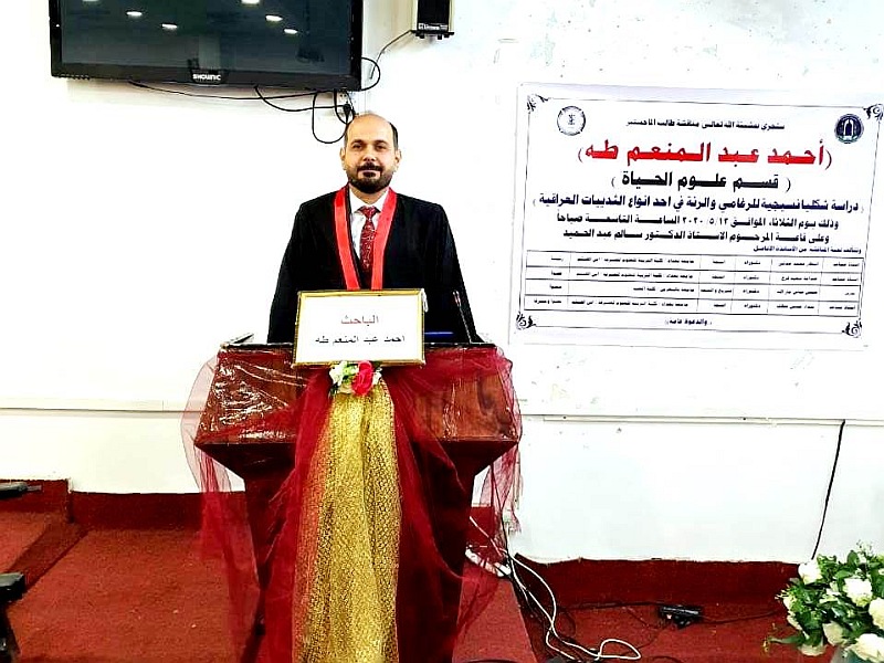
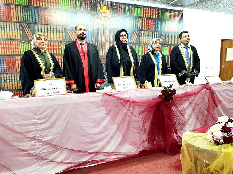
ناقش قسم علوم الحياة في كلية التربية للعلوم الصرفة (ابن الهيثم) رسالة الماجستير الموسومة (دراسة شكليائية ونسجية للرغامى والشجرة القصبية والرئة في بيز ابن عرس العراقي Herpestes javanicus ) للطالب ( احمد عبد المنعم طه الجوادي ) التي انجزها تحت اشراف التدريسية في القسم ( أ. م د. بيداء حسين مطلك ) ونوقشت الرسالة من قبل لجنة المناقشة التي تألفت من الاعضاء المدرجة اسمائهم فيما يأتي :
-
أ.م.د. انتظار محمد مناتي ( رئيسا )
-
أ.م.د. صدامة سعيد فرج ( عضوا )
-
م.د. حسن عباس جار الله ( عضوا )
-
أ.م.د. بيداء حسين مطلك ( عضوا و مشرفا )
وهدفت هذه الدراسة الى التعرف على الوصف الشكليائي والتركيب النسجي للرغامى والشجرة القصبية والرئة لبيز ابن عرس Herpestes javanicus كأحد الثدييات العراقية التي تقطن البيئة العراقية .
استعملت في الدراسة الحالية 15 عينة من الذكور والاناث من بيز ابن عرس يتراوح معدل اوزانها بين288- 564غم , تم استئصال الرغامى والرئة من العينات وثبتت باستعمال المحاليل المثبتة واجريت عليها سلسلة من التحضيرات النسجية وتم استعمال الملون الروتيني الهيماتوكسلين والايوسين وعدد من الملونات الخاصة كملون فان كيزن وملون شف حامض الدوري , واجريت الفحوصات المختبرية على 5عينات من الدراسة للتعرف على تفرعات الشجرة القصيبية باستعمال تقنية صب الراتنج ومن ثم تعريض العينات للتأكل.
أوضحت الدراسة التشريحية للرغامى في ابن عرس بانه يتمثل بأنبوب غضروفي مجوف مرن غير قابل للانطواء، يمتد ذنبيا من نهاية الغضروف الحلقي للحنجرة عند مستوى الفقرة العنقية الثانية او الثالثة , عند منطقة ارتباط الغدة الدرقية بالسطح البطني للحلقات الغضروفية الثلاث الاولى, فضلا عن ان الرغامى يرتبط مع القلب في مستوى قاعدته وعند مستوى الضلع الصدري الخامس عند قاعدة القلب. يتكون الرغامى من حلقات غضروفية غير متفرعة وغير كاملة تشبه شكل حرف C تكون مغلقة من الجانب البطني ومفتوحة من الجانب الظهري وترتبط نهايتي الحلقات الغضروفية بنسيج ضام مفكك والياف عضلية ملساء متمثلة بالعضلة الرغامية ويتراوح عدد الحلقات الغضروفية ما بين 46-47 حلقة غضروفية والتي تم عدها باستعمال ملون ازرق المثلين , يُقسم الرغامى على ثلاثة أجزاء متمثلة بالجزء العلوي والجزء الوسطي والجزء السفلي.
بلغ معدل قطر الجزء العلوي من الرغامى 0.3±4.5 ملم اذ كان اكبر قطراً من معدل الوسطي والجزء السفلي ، في حين بلغ معدل قطر الجزء الوسطي للرغامى0±4 ملم ، اما معدل قطر الجزء السفلي للرغامى فهو اصغر من معدل قطر الجزء العلوي والوسطي اذ بلغ0.3±3 ملم .
أظهرت نتائج الدراسة النسجية للرغامى انه يتألف نسجيا من ثلاث غلالات وطبقة واحدة متمثلة بالغلالة المخاطية والغلالة تحت المخاطية وطبقة من الغضروف الزجاجي والغلالة البرانية تتكون الغلالة المخاطية من البطانة الظهارية والمتمثلة بنسيج ظهاري عمودي مطبق كاذب مهدب حاوٍ على ثلاثة أنواع من الخلايا هي الخلايا العمودية طويلة المهدبة والخلايا والقاعدية وتنتشر بين هذين النوعين الخلايا الكاسية وتستند جميع الخلايا الى الغشاء القاعدي. يستند النسيج الظهاري الى طبقة الصفيحة الأصيلة المكونة من نسيج ضام مفكك اما الطبقة العضلية المخاطية فتكون ضعيفة التكوين ومكونة من الياف عضلية ملساء مبعثرة. بخصوص الغلالة تحت المخاطية فهي تحوي على غدد مصلية و مخاطية وغدد مختلطة, في حين ان الغلالة البرانية تحتوي على نسيج ضام مفكك حاوٍ على اوعية دموية واعصاب , اما الطبقة الغضروفية فتحتوي على الغضروف الزجاجي المرتب على شكل حلقات غير كاملة تشبه حرفC.
يمتلك بيز ابن عرس زوجا من الرئات الاسفنجية ذات اللون الوردي البراق التي تشغل معظم التجويف الصدري . تبدو الرئتان مشابه لورقة البرسيم لها قمة وثلاثة سطوح متمثلة بالسطح الضلعي الذي يكون محدباً, وسطح وسطي, ويمتاز بكونه ضيقا وسطح حجاب حاجزي ويكون مطابقاً للحجاب الحاجز.
أظهرت نتائج الدراسة الحالية ان الرئة اليمنى تكون اكبر حجماً من الرئة اليسرى وتتكون الرئة اليمنى من أربعة فصوص وهي : الفص القمي والفص الوسطي والفص الذنبي وفص مساعد ,
اما الرئة اليسرى فتتكون من ثلاثة فصوص وهي : الفص القمي والفص الوسطي والفص الذنبي, ظهرت قمة الرئة اليمنى صغيرة وشبة حادة اما الرئة اليسرى فتكون مستديرة وعمياء.
بينت الدراسة الحالية لبيز ابن عرس ان الرغامى تتفرع الى فرعين من القصبات الهوائية الاولية (الرئيسية) التي تكون مشابهة في تركيبها النسجي للرغامى , تتفرع القصبات الاولية لتكون القصبات داخل رئوية الثانوية والثالثية إذ ان الحلقات الغضروفية تتحول الى صفائح من الغضروف الزجاجي اصبعي الشكل تختفي هذه الصفائح الغضروفية في القصيبات والتي تتفرع بدورها الى القصيبات النهائية والتي تعطي تفرعاً الى القصيبات التنفسية في المقابل كل تفرع من القصيبات التنفسية يعطي تفرعاً الى عدد من القنوات السنخية التي تفتح في عدد من الاكياس السنخية التي تحوي جدرانها على عدد كبير من الاسناخ الرئوية . اظهرت النتائج الاحصائية ان القصبات الاولية كانت اكبر قطرا من الاقطار الاخرى جاءت بعدها القصبات الثانوية ثم القصبات الثالثية فالقصيبات والقصيبات النهائية التي لم يكن بين اقطارها فرق معنوي بعدها القصيبات التنفسية والقنوات السنخية والاكياس السنخية واخيراً الاسناخ.
أظهرت نتائج الدراسة الحالية ان القصيبات تكون بشكل مقاطع كروية او بيضوية وتتكون جدرانها من طبقة مخاطية مكونة من خلايا النسيج الظهاري العمودي البسيط المهدب الى نسيج ظهاري مكعبي بسيط مهدب في حين كلا النوعين من القصيبات النهائية والتنفسية تبطن بنسيج ظهاري مكعبي واطئ يكون خالي من الخلايا الكاسية وينعدم وجود الغضروف الزجاجي وتصبح الغدد الرغامية قليلة العدد . وتظهر الاكياس السنخية كتراكيب كيسيه غير منتظمة الشكل فضلا عن انها رقيقة الجدران اذ تبطن بخلايا حرشفية بسيطة , اما الكيس السنخي فيتألف بدوره من عدد كبير من الاسناخ الرئوية والتي تظهر بشكل غرف هوائية غير منتظمة صغيرة الحجم وتبطن الاسناخ الرئوية بنوعين من الخلايا :خلايا سنخيه Type I , وخلايا سنخيه Type II.
وخلص الطالب الى التوصيات الاتية :
-
دراسة التزويد العصبي والتزويد الدموي للجهاز التنفسي في بيز ابن عرس.
-
اجراء دراسات مقارنة لأنواع مختلفة من الثدييات العراقية المختلفة المعيشة .
-
اجراء دراسة نسجيه للرئة باستعمال المجهر الالكتروني
Electron microscope للتعرف على التركيب المستدق للخلايا والاهداب بصورة اكثر تفصيلا.
-
اجراء دراسة كيمو نسجية مناعية للتعرف على وظيفة الخلايا الغبارية Dust cells .
-
اجراء دراسة كيمو مناعية نسجية للتعرف على خلايا الصم العصبية Neroendocrine cells.
-
اجراء دراسات حول التكوين الجنيني لرغامى ورئة بيز ابن عرس.
Morphological and Histological Study of Trachea , Bronchial tree and Lung in Iraqi Weasel
(Herpestes javanicus)
By Ahmed .A.Taha
Supervised by Assit. Prof. Dr. Baydaa Hussain Mutlak
Summary
The current study aimedidentify the Morphometric description and histological composition of the trachea, bronchial tree and lung of Weasel Herpestes javanicus as one of the Iraqi mammals inhabiting the Iraqi environment.
A samples of 15 male and female were used from Weasel, with an average weight of 288 and 564 gm were included in the study. Both Trachea and lung were removed as one piece from the samples and they were fixed using fixative solutionsand conducted a series of serialtissue preparations steps as well as using routinestain hematoxylin & eosin and a number of special stains such as Van Gieson’s and periodic acid-Schiff (PAS) stains.The laboratory tests were carried out on the bronchial tree using the technique of casting resin and then exposing it tocorrosion on 5samples.
The anatomical study of the tracheal in Weasel explained that it was consists of a flexible, hollow, cartilage tube that was non- folding. Which extends from the end of the cricoid cartilage of the larynx at the level of the second or third cervical vertebra, at the area of the thyroid gland with the abdominal surface of the first three cartilaginous tracheal , Caudal part of the trachea is related to the base of the heart at the level of the fifth thoracic rib at the. The tracheal consists of non-branched and incomplete cartilaginous rings resembling the shape of the letter C, closed from the ventral side and open from the dorsal side. The end two of the rings is connected by the tracheal muscles .The number of the tracheal rings ranging from 46-47 rings counted by used the Methylene blue stain, the trachea is divided into three parts represented by( the upper, the middle and the lower) parts . The mean diameter of the upper part of the trachea was 0.3 ± 4.5 mm, while the diameter of the middle and the lower part showed mean of 0 ±4 mm and0.3 ± 3 mm respectively
The results of the histological study of trachea showed that it consisted histologically of three tunics and one layer represented by the tunica mucosa ,tunica submucosa, tunica adventitia and cartilage layer. The tunica mucosa consists of the epithelial lining, which is represented by a ciliated pseudostratified columnar epithelial tissue that has three types of cells, ciliated columnar cells, basal cells, and goblet cells are spread between these two types, and all cells are based on the basal membrane. The epithelial tissue is supported by the lamina propria , which is made up of loose connective tissue ,while the Muscular is mucosa layer is consists of scattered smooth muscle fibers
Concerning the subcutaneous mucosa, it contains serous and mucous glands and mixed secretory glands, while the tunica adventitious contains a loose connective tissue containing blood vessels and nerves, and the cartilage layer contains the hyaline cartilage arranged in the form of incomplete rings resembling the letter C
The weasel has a pair of bright pink spongy lungs that occupy most of the thoracic cavity. The lungs look similar to Clover leaf pattern having a apex and three surfaces represented by the costal surface which is convex, externally the mediastinal surface , which is characterized by narrow concave shape and the diaphragmatic surface that related to the diaphragm.
The results of the current study showed that the right lung is larger than the left lung and the right lung consists of four lobes: the apical ,middle, caudal and the accessary lobe ,while the left lung consists of three, which are:( the apical, middle, and caudal lobe). Theapex of the right lung looks small and semi-sharp. The left lung is round and blunt. The current study of Weas showed that the tracheal branched into two features of the primary (main) bronchi (Right&Left) , which looks similar in their histological to that of the trachea, the bronchi divided in secondary and tertiary intra pulmonary bronchi ,as the cartilage rings turn into sheets of the hyaline cartilage that disappeared in the bronchioles , which in turn branched into terminal bronchioles, that gives origin to the respiratory bronchioles, which in contrast each one of therespiratory bronchiolesgives to a number of alveolar ducts that open in several alveolar sacs whose walls contain a large number of pulmonary alveoli.
Statistical results showed that the primary bronchi was larger indiameter than of the other , followed by the secondary bronchi, then the tertiary bronchi, bronchioles,and terminal bronchioles , which did not have a significant difference between their dimensions, then the respiratory bronchioles, alveolar tracts, alveolar sacs, and finally alveoli.
The results of the current study showed that the bronchioles are in the form of spherical or oval sections and their walls consist of a mucous layer composed of ciliated simple columnar epithelial tissue cells to simple ciliated cuboidal epithelial tissue while both types of terminal and respiratory bronchioles are lined with low cuboidal epithelial tissue that is free of goblet cells and the hyaline cartilage is absent. The presence of the endotracheal gland become few. The alveolar sacs appear as irregular sacs structures as well as thin-walled as they line with simple squamous cells. The alveolar sac, in turn, consists of a large number of pulmonary alveoli that appear as irregular and small air chambers. The alveolar sac, in turn, consists of a large number of pulmonary alveoli that appear as small, irregular air chambers and line the pulmonary alveoli with two types of cells: alveolar cells Type I, and cells type II.