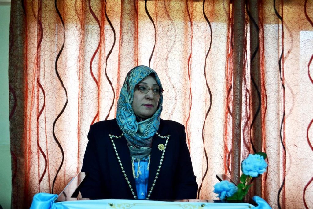
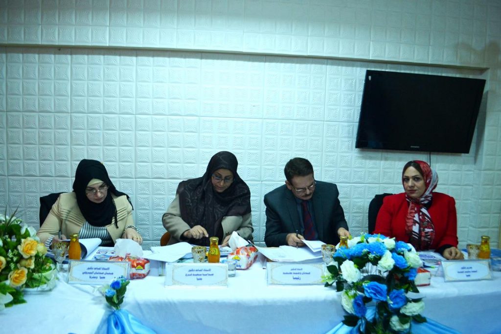
جرت يوم الاحد الموافق 23/ 12/ 2018  على  قاعة الندوات في قسم علوم الحياة المناقشة العلنية لرسالة طالبة الماجستير ماجدة جمعة راضي السراجي الموسومة :

دراسة نسجية مرضية لخصى وبرابخ الفئران البيض المعاملة بأوكسيد الزنك النانوي


حيث تألفت لجنة المناقشة من التدريسيين الأفاضل :

أ.د . عدنان وحيد محمد                    رئيسا

أ.م.د . صدامة سعيد فرج                 عضوا

أ.م.د .اسماء بشير عبد                    عضوا

ا.م.د .جنان عدنان البيروتي             عضوا ومشرفا

اجريت الدراسة على 30 فاراً ابيض سويسرياً التي تم الحصول عليها من هيأة البحث والتطوير ، بهدف معرفة تأثير جسيمات اوكسيد الزنك النانوي (ZnO – NPs) في بعض اعضاء الجهاز التكاثري والغدد الملحقة به لذكور الفئران البيض. جرعت الحيوانات بجسيمات اوكسيد الزنك النانوي عن طريق الفم بجرعة بلغت 0.1 مل وبتركيز 100 و200 ملغم/كغم وللمدد 7 و14 يوماً وبعدها تم تشريح الحيوانات في اليوم التالي من انتهاء مدتي التجريع واخذت منها الخصى والبرابخ وبعض الغدد الملحقة (الحويصلة المنوية وغدد كوبر) لدراسة المعايير المظهرية والتشريحية التي ظهرت نتيجة المعاملة بجسيمات المادة النانوية وهذه المعايير شملت الاتي:-

1-         اوزان الحيوانات قبل وبعد المعاملة بـ ZnO-NPs واوزان الخصى ورؤوس وذيول البرابخ واوزان الحويصلة المنوية وغدد كوبر.

2-         التغيرات في مواصفات النطف في ذيول البرابخ والتي تمثلت بحساب النسبة المئوية للتشوهات النطفية.

3-         التغيرات الحاصلة في اقطار النبيبات المنوية وسمك الجدار المنوي للنبيبات المنوية في الخصى.

4-         التغيرات الحاصلة في اعداد خلايا لايدج واقطار أنويتها.

5-         التغيرات الحاصلة في سمك الجدار وارتفاع الظهارة المبطنة لكل من رأس وذيل البربخ.

6-         التغيرات الحاصلة في ارتفاع الظهارة واقطار انوية الخلايا للحويصلات المنوية.

7-         التغيرات الحاصلة في اقطار الحويصلات العنبية واقطار الخلايا العنبية وانويتها لغدد كوبر.

8-         التغيرات المرضية النسجية للخصى والبرابخ (الرأس والذيل) والحويصلات المنوية وغدد كوبر باستعمال صبغتي الـPAS  و الهيماتوكسلين–ايوسين.

اظهرت الملاحظات المظهرية حدوث تغيرات تمثلت بقلة المادة الدهنية المحيطة بالخصى واحتقان الاوعية الدموية في الذكور المعاملة بتركيزي 100) و 200 ملغم/كغم من ZnO-NPs) اذ كانت هذه التغيرات واضحة في الخصى خلال مدة المعاملة  14يوماً مقارنة بحيوانات السيطرة. وسجلت نتائج الدراسة انخفاضاً معنوياً(P<0.05)  في متوسط اوزان اجسام الحيوانات والخصى ورؤوس وذيول البرابخ مقارنة مع السيطرة وقد ازداد الانخفاض بزيادة التركيز ومدة المعاملة. في حين سجلت الدراسة حصول زيادة معنوية (P<0.05) في متوسط اوزان الحويصلات المنوية وغدد كوبر للحيوانات المعاملة مقارنة مع مجموعة السيطرة وازدادت اوزان هذه الاعضاء بزيادة التركيز وزيادة مدة المعاملة.

بينت نتائج الدراسة حصول زيادة معنوية (P<0.05) في النسبة المئوية للتشوهات النطفية في ذيول برابخ الحيوانات المعاملة بـ 100 و 200 ملغم/كغم من ZnO-NPs وللمدتين 7 و14 يوماً عند مقارنتها مع حيوانات السيطرة ، وكانت نسبتها متزايدة بزيادة التركيز ومدة المعاملة ومن هذه التشوهات انعدام رأس النطفة والتواء الرأس وظهور القطرة الهيولية في القطعة الوسطية وانحناء الذيل والتفافه.

سجلت الدراسة ايضاً حصول انخفاضاً معنوياً (P<0.05) في اقطار النبيبات المنوية وسمك الجدار المنوي للخصى ، فضلاً عن حصول انخفاض في سمك الجدار وارتفاع الظهارة لكل من رؤوس وذيول البرابخ في الحيوانات المعاملة بالتركيزين ومدتي التعرض 7 و 14 يوماً وكان الانخفاض متزايداً بزيادة التركيز ومدة التعرض. اما اعداد خلايا لايدج واقطار أنويتها فقد اظهرت الدراسة حصول زيادة معنوية (P<0.05) في اعداد الخلايا وانخفاضاً معنوياً (P<0.05) في اقطار أنويتها لكلا التركيزين ومدتي المعاملة وان اعلى تغير قد لوحظ في التركيزـ 200 ملغم/كغم من ZnO-NPs  والمدة 14 يوماً عند مقارنتها مع حيوانات السيطرة.

 كما اظهرت النتائج حصول اضرار نسجية في الخصى تمثلت بحدوث انخفاض في سمك النسيج القاعدي وتفجي سايتوبلازم الخلايا المبطنة للنبيب المنوي وتنخر بعض خلايا النبيبات المنوية وتضرر الالياف المحيطة بالغشاء القاعدي للنبيب المنوي. فضلا عن حصول اضرار في رؤوس وذيول البرابخ تمثلت بتحطم الخلايا الظهارية وانفصال الظهارة عن الغشاء القاعدي وقلة اعداد النطف واتساع الفسح بين النبيبات البربخية  وقلة النسج الضامة فضلا عن فرط التنسج للظهارة المبطنة للنبيبات و ضمور انوية الخلايا الظهارية وكذلك ظهور الخلايا الالتهامية في تجويف  نبيب ذيول البرابخ مع احتقان الاوعية الدموية.

اظهرت المعاملة بـ ZnO-NPs اضرارا نسجية في الحويصلة المنوية تمثلت بقلة الافرازات الحويصلية في تجاويف الحويصلة و تفجي سايتوبلازم الخلايا الظهارية و تغلظ انوية الخلايا و زيادة ارتفاع الطيات الحويصلية . اما غدد كوبر فقد تضررت هي الاخرى من خلال تحطم بعض الخلايا الظهارية للعنيبة وظهور الحبيبات الافرازية وتراكم الخلايا الالتهابية ما بين الحويصلات العنيبية واحتقان الوعاء الدموي وزيادة الفسح ما بين الحويصلات العنيبية ونسج السدى للحيوانات المعاملة بـ ZnO-NPs ولكلا مدتي التعرض مقارنة بمجموعة السيطرة. ونتيجة للمعطيات التي تم الاشارة اليها في هذه الدراسة يتبين ان جسيمات اوكسيد الزنك النانوية وعلى الرغم من استعمالاتها الواسعة والمتعددة الا انها يمكن ان يكون لها تأثيرات سلبية وضارة لاسيما في الجهاز التكاثري لذكور الفئران البيض.

وحضر المناقشةجمع من التدريسيين وطلبة الدراسات العليا وذوي الطالبة والضيوف الكرام .تمنياتنا للطالبة الموفقية والنجاح.


M.Sc. Thesis Discussion of student (Majda Juma’a Radhi Al-Saraji) from department of Biology

On Sunday, the 23th of December 2018, at the hall of seminar in the Biology department, M.Sc. Thesis Discussion of student (Majda Juma’a Radhi Al-Saraji) from department of Biology entitled (Histopathological Study in testes and epididymis of albino mice treated with Zinc oxide nano particles) has been discussed

The discussion committee

Prof.Dr.Adnan Waheed Mohammed      head

Asst.prof.Dr.Sadamah Said Faraj    member

Asst.prof.Dr.Asma’a Basheer Abid    member

Asst.prof.Dr.Jinan Adnan Al-Bairooti    supervisor member

The Abstract

The study was conducted on 30 Swiss albino mice, in order to determine the effect of ZnO-NPs on some reproductive organs and accessory glands of male albino mice. Animals were oral administrated with 0.1 ml of 100 and 200 mg/kg ZnO nanoparticles for 7 and 14 days. Animals were then sacrificed in the next day after the end of the two dosing period and took from them testes, epididymis, and some accessory glands (seminal vesicle and Cowper’s glands) to study the morphological and anatomical criteria that appeared as a result to treated with ZnO-NPs. These criteria as followed

. Animal weights before and after treatment with ZnO-NPs, weights of testes, head and tails of epididymis, weights of seminal vesicle and Cowper glands

– Changes in sperm characteristic in the epididymis tail that involved accounted the percentage of sperm abnormalities

– Changes in the diameter of seminiferous tubules and the wall thickness of seminiferous tubule in the testis

– Changes in the number of leydig cells and their nuclei.

– Changes in the wall thickness and lining epithelium heightin the head and tail of the epididymis.

-Changes in the epithelium height and the diameter of cells nuclei in the seminal vesicles.

– Changes in the diameters of the acini vesicles and the diameters of the acini cells and their nucleiin the Cowper glands.

. Histopathological changes in the testes, epididymis (head and tail), the seminal vesicles and the Cowper glands by using PAS and hematoxylin-eosin stains.

The visual observations showed a decrease in the amount of lipid surrounding the testis and blood vessels congestion in the male treated with 100 and 200 mg / kg of ZnO-NPs. These changes were evident in the testis during the period of 14 days compared to control animals. The results of this study showed a significant decrease (P <0.05) in the average weights of animals body, testes, head and tails of epididymis compared with the control. The decreasing weight was increased by increasing the concentration and duration of treatment. The study showed a significant increase (P <0.05) in the average weight of seminal vesicles and Cowper glands of treated animals compared with the control group and the weight of these organs were increased with increasing the concentration and duration of treatment. The results of this study showed a significant increase (P <0.05) in the percentage of sperm abnormalities in epididymis tail of animal treated with 100 and 200 mg / kg of ZnO-NPs for 7 and 14 days when compared with control animals. These abnormalities were increased by increasing the concentration and duration of treatment.The sperm abnormalities involved the absence of sperm head, head twist, appearance of cytoplasmic droplet in the mid piece, bend and twist of sperm tail. The study also showed a significant decrease (P <0.05) in the diameter of the seminiferous tubules and the thickness of the seminiferous wall in the testes. There was also a decrease in the wall thickness and the height of epithelium in both head and tail of the epididymis in animals treated with two concentrations and two duration of exposure 7 and 14 days. These decline was increasing with increased the concentration and duration of treatment. The results showed a significant increase (P <0.05) in the number of leydig cells and a significant decrease (P <0.05) in thediameter ofleydig cell nuclei with both concentrations and duration of treatments. The highest change was observed in the concentration of 200 mg / kg of ZnO-NPs and duration 14 days when compared with control animals .The results also showed injuries in the tissues of testis, which involved a decrease in the thickness of the basal tissue, cytoplasmic vacuolation in lining epithelium of seminiferous tubule, cells necrosis in some seminiferous tubule, and fibers damaging that surrounding the basement membrane of the seminiferous tubules, as well as induce damage in the tissues of head and tail of epididymis which involved the destruction of epithelial cells, the epithelium separation from the basement membrane ,decline of sperm number, widening of space between epididymis tubules, and the decline of connective tissue as well as hyperplasia of lining epithelium of the tubules and epithelial cell nuclei atrophy in addition to the appearance of inflammatory cells in the tubular cavity of epididymis tail with blood vessel congestion. The treatment with ZnO-NPs showed injuries in the tissue of seminal vesicles, consisting of the decline of vesicular secretions in the vesicle cavities, cytoplasmic vacuoles of epithelial cells, pyknosis of cell nuclei, increased vesicle fold height. Cowper’s glands were also damaged by the destruction of some epithelial cells in the acini, the appearance of secretory granules, the accumulation of inflammatory cells between the acini vesicles, blood vessel congestion and increasing the space between the acini vesicles and stroma tissues in animals treated with ZnO-NPs with both duration of exposure compared to the control group. As a result of the data referred to in this study, the zinc oxide nano particles, despite their extensive and multiple uses, but can have adverse effects and harmful, especially in the reproductive system of male albino mice.

كلية التربية للعلوم الصرفة ابن الهيثمAuthor posts

Avatar for كلية التربية للعلوم الصرفة ابن الهيثم

كلية التربية للعلوم الصرفة (ابن الهيثم) - College of Education for Pure Science (Ibn Al-Haitham)

Comments are disabled.