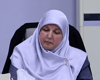
ناقش قسم علوم الحياة في كلية التربية للعلوم الصرفة ( ابن الهيثم ) رسالة الماجستير الموسومة ( دراسة شكليائية وكيمونسجية لاجزاء المعي الدقيق في السنجاب القوقازي Sciurus anomalus ) للطالب ( أحمد مصطفى أحمد ) التي انجزها تحت اشراف التدريسية في القسم ( أ.م.د. انتظار محمد مناتي ) ونوقشت من قبل لجنة المناقشة التي تألفت من الاعضاء المبينة اسمائهم فيما يأتي :
- أ.م.د. ايمان سامي احمد (رئيسا)
- أ.م.د. هدى رشيد كريم (عضوا)
- أ.م.د. بيداء حسين مطلك (عضوا)
- أ.م.د. انتظار محمد مناتي (عضوا و مشرفا)
وقد بينت الطالبة الباحثة ان بحثها هذا جاء نتيجة قلة الدراسات والبحوث عن الوصف الشكليائي والتركيب النسجي للمعي الدقيق في الفقريات البرية العراقية , اذ نالت الطيور والحيوانات المختبرية أهتمام الباحثين , في حين ان الفقريات البرية لاسيما الثدييات البرية لم تحض بنصيب من تلك الدراسات , بالرغم من امتلاك العراق من شماله الى جنوبه بتنوع بيئي, جعل العديد من الحيوانات البرية المتنوعة تتواجد في بيئته ,هذا شكل حافزاً لدراسة المعي الدقيق كأحد اجزاء القناة الهضمية والتعمق بتفاصيله الدقيقة , اذ لا توجد دراسات تخص موضوع الدراسة الحالية ضمن المصادر المتوفرة, وقد تم اختيار أحد انواع الثدييات البرية العراقية المتمثلة بالسنجاب القوقازي Sciurus anomalus , وتم اجراء الدراسة الحالية للتعرف على الوصف الشكليائي والتركيب النسجي فضلاً عن دراسة القياسات الشكليائية والنسجية لاجزاء المعي الدقيق كافة المتمثلة بالعفج Duodenum والصائم Jejunum واللفائفيIleum
وصممت الدراسة الحالية للتعرف على الوصف الشكليائي والتركيب النسجي للمعي الدقيق في السنجاب القوقازي Sciurus anomalus وتضمنت هذه الدراسة القياسات الشكليائية والقياسات النسجية المترية فضلاً عن الدراسة النسجية والكيمونسجية للأجزاء الثلاثة للمعي الدقيق ، حيث استعمل في الدراسة الحالية 15عينة من السناجب الحية البالغة, التي تم تخديرها بمادة الكلوروفورم وتم قياس كل من طول الجسم الحقيقي وطول الجسم القياسي ثم تم التضحية بالحيوانات واستئصل منها المعي الدقيق وتم قياس اطوال واوزان المعي الدقيق واجزائه الثلاثة (العفج والصائم واللفائفي) ثم ثبتت العينات بمحاليل التثبيت واجريت عليها الخطوات المتسلسلة في تحضير الشرائح النسجية ثم لونت باستعمال الملونات الروتينية والخاصة (ملون الهيماتوكسلين والايوسين, ملون شف الحامض الدوري, ملون الاليشين الازرق, ملون فان كيزن, ملون ماسون ثلاثي الكروم) وطريقتي التلوين (سنكا و كريملز )واظهرت نتائج الدراسة الحالية ماياتي:-
-
قياسات الدراسة الشكليائية: بينت نتائج الدراسة الاحصائية وجود علاقة ايجابية بين معدل طول الجسم الكلي وطول المعي الدقيق فضلاً عن معدل اطوال واوزان الاجزاء الثلاث للمعي الدقيق عند مستوى احتمالية P .
-
الوصف الشكليائي : يعد المعي الدقيق الجزء الاكثر طولاً في القناة الهضمية ويبدو بهيأة التواءات او التفافات كثيرة غير منتظمة الشكل, يقع المعي الدقيق في المنطقة الوسطى من التجويف البطني بين المعدة والجزء الاول من المعي الغليظ المتمثل بالاعور , يبدأ المعي الدقيق من البواب المعدي وينتهي عند الصمام اللفائفي الاعوري ويقسم على ثلاثة اجزاء رئيسة متمثلة بالعفج والصائم واللفائفي.
-
يمثل العفج الجزء الاول من المعي الدقيق ويبدأ من نهاية المعدة تحديداً من البواب المعدي ويستمر حتى نهاية الذراع الصاعد للعفج ويتصل بالصائم عن طريق الانثناء العفجي الصائمي, ويكون بشكل حرف C حول البنكرياس, يقسم العفج في السنجاب القوقازي موضوع الدراسة الحالية على خمسة اجزاء متمثلة بالعفج القحفي والانثناء العفجي القحفي والعفج النازل والانثناء العفجي الذنبي والعفج الصاعد.
-
يمثل الصائم اطول جزء في المعي الدقيق ويبدأ من الانثناء العفجي الصائمي وينتهي عند بداية الطية اللفائفية الاعورية, يقع الصائم في الجزء المركزي من التجويف البطني بين المعدة قحفياً والاعور والمثانة البولية ظهرياً , ويظهر الصائم بشكل لفائف عديدة تأخذ بالانخفاض تدريجياً عند اقترابنا من بداية اللفائفي وتشكل شكل يشبه باقة الورد عند اتصالها بالمساريق .
-
يعد اللفائفي الجزء النهائي والاخير للمعي الدقيق ويأتي بعد العفج والصائم ويوصف بكونه انبوباً قصيراً مستقيماً خالٍ من الالتفافات العديدة , ويقع اللفائفي في المنطقة الوسطى من التجويف البطني داخل تقعر الاعور, ويبدأ من بداية الطية اللفائفية الاعورية وينتهي عند الصمام اللفائفي الاعوري.
-
تتصف البطانة الداخلية للعفج والصائم بوجود عدد من البروزات الكثيفة والمرتفعة تدعى بالزغابات الا انها في اللفائفي تكون اقل كثافة وطولاً من نظيرتها في العفج والصائم فضلاً عن وجود عدد من الطيات او ماتعرف بصمامات كيركرنك في الاجزاء الثلاثة للمعي الدقيق, اذ ظهرت الطيات الدائرية في الجزء القحفي للعفج , فيما برزت الطيات الطولية في الجزء النازل والصاعد منه, في حين ظهرت الطيات الطولية في الجزء الامامي للصائم, اما في اللفائفي فظهرت الطيات الطولية في الجزء الامامي والوسطي من تجويفه , في حين كانت مفقودة في الجزء الخلفي منه.
-
التركيب النسجي : اظهرت نتائج الدراسة الحالية ان جدار المعي الدقيق يتالف نسجياً من اربع غلالات رئيسة متمثلة بالغلالة المخاطية وتحت المخاطية والعضلية والمصلية.
-
تتالف الغلالة المخاطية من ثلاث طبقات ثانوية تتضمن طبقة البطانة الظهارية والصفيحة الاصيلة و الطبقة العضلية المخاطية , تتحور البطانة الظهارية الى العديد من الزغابات ويغلف السطح الخارجي للزغابات بنسيج ظهاري عمودي بسيط , وتتباين اشكال واطوال وكثافة الزغابات في الاجزاء الثلاثة للمعي الدقيق , اذ تميزت زغابات العفج بكونها ورقية الشكل, بينما يظهر القليل منها شكلاً صولجانياً, في حين اتصفت زغابات الصائم بكونها ورقية الشكل ذات نهايات مدببة او مدورة سميكة , في حين يظهر البعض منها شكلاً اصبعياً, بينما في اللفائفي تكون الزغابات اقل كثافة وطولاً فقد تبدو ورقية الشكل وقصيرة, في حين يظهر البعض منها شكلاً اسطوانياً وصولجانياً.
-
تتالف الصفيحة الاصيلة في الاجزاء الثلاثة للمعي الدقيق من نسيج ضام مفكك, وتقع الغدد المعوية فيها وتكون الغدد من النوع النبيبي المستقيم البسيط , والخلايا المكونة لهذه الغدد هي خلايا عمودية وخلايا كأسية وخلايا غير متمايزة (خلايا جذعية) وخلايا بانيث فضلاً عن وجود خلايا الصم المعوية .
-
اظهرت خلايا الصم المعوية بنوعيها (الاليفة والمحبة للفضة) في النسيج الظهاري المغلف للزغابات والمبطن للغدد المعوية في الأجزاء الثلاث للمعي الدقيق تفاعلاً ايجابياً معتدلاً عند استعمال طريقتي التلوين(سنكا و كريملز) .
-
تتألف الطبقة العضلية المخاطية في العفج والصائم واللفائفي من طبقة رقيقة جداً ومفردة من الألياف العضلية الملساء المرتبة دائرياً.
-
تتالف الغلالة تحت المخاطية في الاجزاء الثلاثة للمعي الدقيق من نسيج ضام كثيف, وتمتاز بوجود غدد برونر والمتواجدة في العفج فقط والتي تكون من النوع النبيبي الحويصلي المركب, بينما يمتاز جزأي الصائم واللفائفي بوجود عقيدات لمفية متجمعة تدعى بلطخ باير.
-
اظهرت الوحدات الافرازية لغدد برونر تفاعلاً ايجابياً قوياً عند تلوينها بملون الاليشين الازرق, في حين اظهرت الوحدات الافرازية لهذه الغدد تفاعلا ايجابيا معتدلا عند تلوينها بملون PAS.
-
تتالف الغلالة العضلية في الاجزاء الثلاثة للمعي الدقيق من طبقتين ثانويتين من الالياف العضلية الملساء , الداخلية دائرية الترتيب وسميكة, والخارجية طولية الترتيب واقل سمكاً, ويفصل بين الطبقتين نسيج ضام ليفي يحتوي على ضفائر اورباخ .
-
تتالف الغلالة المصلية في الاجزاء الثلاثة للمعي الدقيق من نسيج ضام مفكك , ويحدها من الخارج صف من الخلايا المتوسطة.
-
القياسات النسجية المترية
-
بينت نتائج الدراسة الاحصائية ان معدل ارتفاع الزغابات في الاجزاء الثلاثة للمعي الدقيق يأخذ بالتناقص التدريجي من العفج باتجاه اللفائفي. ان معدل طول الخبايا يأخذ بالتناقص التدريجي من العفج باتجاه اللفائفي وسجلت اعلى قيمة له في كل من العفج والصائم, اما عرض الخبايا فقد كانت اكثر عرضاً مقارنة باللفائفي.
-
يزداد معدل عدد الخلايا الكأسية في جزء اللفائفي ويقل تدريجياً باتجاه العفج, في حين كان على العكس من ذلك اذ ان معدل عدد خلايا بانيث في جزء العفج والصائم اعلى مما هو عليه في اللفائفي.
-
يزاد معدل عدد غدد برونر في الجزء القحفي للعفج ثم تأخذ بالتناقص التدريجي في الانثناء العفجي القحفي وبلغت اقل عدداً لها عند الجزء النازل .
-
ان معدل سمك الغلالة المخاطية والغلالة تحت المخاطية والغلالة المصلية كانت اكبر في جزء العفج مقارنة بجزأي الصائم واللفائفي , فيما ظهرت الغلالة العضلية في اللفائفي اكثر سمكاً من مثيلاتها في جزأي العفج والصائم .
ولكون نتائج الدراسة الحالية جوانب معينة وفتحت المجال للتقصي والدراسة عن جوانب اخرى للوصول الى الحقائق العلمية فقد خلصت الطالبة الى التوصيات الاتية:-
1.اجراء دراسة نسجية مقارنة للمعي الدقيق مع انواع اخرى من الثدييات البرية التي تعيش في البيئة العراقية ذات تغذية وبيئات مختلفة والتي لم تحضَ بنصيب من الاهتمام العلمي الكافي.
2.اجراء دراسة كيمونسجية للمعي الدقيق للكشف عن الاحماض النووية والدهون .
3.اجراء دراسة تشريحية ونسجية عن التزود العصبي والتجهيز الدموي للمعي الدقيق في السنجاب القوقازي.
4.اجراء دراسة نسجية بأستعمال المجهر الالكتروني الماسحScaning electron microscope للتعرف على انواع واشكال الزغابات في الاجزاء الثلاث للمعي الدقيق.
5.اجراء دراسة شكليائية ونسجية مفصلة عن لطخ باير في انواع مختلفة من الثدييات التي تعيش في البيئة العراقية.
Morphological and Histochemical study of small intestine parts in Caucasian squirrel
(Sciurusanomalus)
By Ahmed .M.Ahmed
Supervised by Assist.Prof.Dr.Intidhar Mohammed Mnati
Abstract
The present study was designed to investigate the morphological, description and histological structure of the small intestine in the Caucasian squirrels)Sciurusanomalus(.Morphological measurements histomorphometric, the histological and histochemical study of the three parts of small intestine have been included
Fifteen samples of adult living squirrels were used, after anesthetization by chloroform, both total body length and real body length were measured. The length and weights three parts of the small intestine (duodenum,Jejunumand ileum) were measured.After that the samples was fixed with fixative solutions and the sequent steps of preparing histological slides were done after they were stained by using routine and special stains (Hematoxlin and Eosin stain, Periodic acidshiff stain,Alcian blue stain, Van gieson’s stain, Masson’strichrome stain) by using two staining methods( Singh’s and Grimelius’ method), the results of the present study showed the following
Measurements of the morphological study
The results of the statistical study showed a positive relationship between the overall body length mean and small intestine length ,as well as the lengths and weights of the three parts of the small intestine at probability level P≤0.05
Morphological description
The small intestine is the longest part of the gut and it appears as a simple pipe with many irregular twists or coils.The small intestine is located in the middle area of the abdominal cavity between the stomach and the first part of the large intestine represented the ceacum.It begins from the pylorus and ends at the ileoceacal valve and divided into three main parts(duodenum , jejunum and ileum
The duodenum represented the first part of the small intestine, begins from the end of the stomach specifically from the Gastric pylorus and continues until the end of the ascending duodenum. It communicates with the jejunum by duodenojejunal flexure, and it forms a C Letter like shape around the pancreas,this duodenum in the Caucasian squirrel subject of the present study divided into five parts,That represented by cranial duodenum, cranial duodenum flexure, descending duodenum, caudal duodenum flexure, and ascending duodenum
The jejunum represented the longest part of the small intestine and begins from the duodenojejunal flexure and it ends at the the beginning of the ileocealfold, the jejunum is located in the central part of the abdominal cavity between the stomach cranially, the ceacum and the urinary bladder dorsally, the jejunum appears in the form of several coils take these coils downwards gradually as we approach the beginning of the ileum and form a shape similar to the festoon when connected to them esentry
The final parts of the small intestine is represented by the ileum ,that comes after the duodenum and jejunum and is described as a short and straight tube and empty of several coils, the ileum is located in the middle area of the abdominal cavity inside the ceacum, and starts from the beginning of the ileoceacal fold and ends at theileoceacal valve
The internallining of the duodenum and jejunum is characterized by the presence of a number of dense and high protrusions called villi.However,the villi in the ileum a reless dense and large than the once in the duodenum and jejunum as well as the presence of a number of folds or what is known as kerckring valves in the three parts of the small intestine, where the folds appeared circular in the cranial part, while longitudinal folds appeared in the descending and ascending part of the duodenum, However the longitudinal folds appeared in the anterior parts of the jejunum, while in the ileum the longitudinal folds appeared in the anterior and median part while they were missing at the posterior part
Histological description
The present study showed that the wall of the small intestine is consisted of four main tunica, represented by tunica mucosa, submucosa, muscularis and serosa
The tunica mucous consists of three sub-layers, including epithelial lining layer, lamina properiaand muscularis mucosa layer, The lining epithelium modified into many villi and the outer surface of the villi encased with a simple columnar epithelial tissue,and variation in the shape, length and density of the villi symbiosis has been observed in the three parts of the small intestine. These variations are clearly characterized in duodenum by a leaf-shaped, while a few of them appear as a mace shape, while the jejunum were characterized by leaf shape, with pointed or rounded ends.Some villi appear as a finger-shaped, while in the ileum the villi are less dense and long, they may look leaf shaped and short, while some of them appear a cylindrical and mace shape
The lamina properia of three parts of the small intestine is composed of Loose connective tissue, the with an intestinal glands that are located in them.These glands are simple straight tubular glands, and the cells that make up these glands are columnar cells, goblet cells, undifferentiated cells (stem cells) and,paneth cells as well as the presence of enteroendocrine cells
The enteroendocrine cells of both types (Argentaffin and Argyrophil) in the epithelial tissue encased of the villi and lining of intestinal gland in the three parts of the small intestine showed the cells a moderate positive when used the two stain (Singh’s and Grimelius’methods
The muscularis mucosa layer of the duodenum , jejunum and ileum consists of a very thin and single layer of smooth muscle fibers that circularly arranged
The tunicasubmucosa of a three parts of small intestine is composed of dense connective tissue, characterized by the presence of Brunner’s glands, and present in duodenum only, which are a type of compound tubulaveolar glands. , while the parts of jejunum and ileum is characterized by the presence of aggregated lymph nodules called Peyers patches
The secretory units of Brunner’s glands showed a strong positive reaction when they stained with Alcian bluestain(AB), while the secretory units of Brunner’s gland showed a moderate positive reaction when stain with PAS
The tunica muscularisof the three parts of the small intestine is consisted of two secondary layers of smooth muscle fiber, thicker inner circular arranged while the outer layer is longitudinal and thinner, these layers separated by fibrous connective tissue containing the auerbach’s plexus
The tunica serosa of the three parts of the small intestine is composed of loose connective tissue, with blood vessels, nerves, and bordered from the outside by a row of mesothelium cells
Histomorphometric measurements
The results of the statistical study showed that the mean of elevation of the villi in the three parts of the small intestine is gradually decreasing from duodenum towards the ileum. that the rate of length of the crypts takes a gradual decrease from duodenum towards the ileum and recorded the highest value in the part of duodenum and jejunum ,In the case ofcrypts width , the results of the study showed that the part of the duodenum and jejunum was more wide compared to the ileum
The mean number of goblet cells in the ileum part increases and gradually decrease towards the duodenum .The mean number of paneth cells in the duodenum and jejunum part is higher than in the ileum
Themean number of Brunner’s glands increase in the cranial part of the duodenum , and then gradually decrease in the duodenum cranial flexure and the showedless number in a descending part
The mean thickness of tunica mucous, submucosa and serosa it was bigger in the part of the duodenum compared to the jejunum and ileum parts, while the tunica muscularis in the ileum appeared to be thicker than other parts of the duodenum and jejunum


