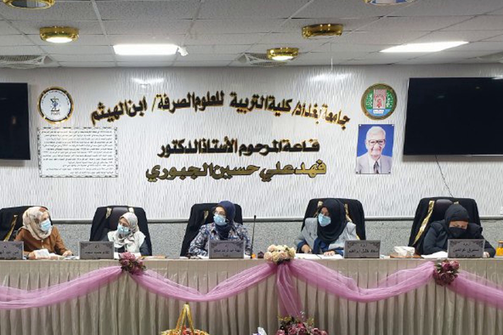
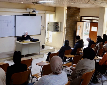
ناقش قسم علوم الحياة في كلية التربية للعلوم الصرفة (ابن الهيثم) رسالة الماجستير الموسومة (دراسة ضراوة بكتريا Cronobacter sakazakii خارج وداخل جسم الكائن الحي) للطالب (حيدر عادل موسى علي) التي انجزها باشراف تدريسيتا القسم (أ.د. لمى عبد الهادي زوين) و (أ.م.د. استبرق عز الدين محمود) ونوقشت من قبل أعضاء لجنة المناقشة المبينة أسمائهم في ما يأتي :
-
أ.د. نهلة عبد الرضا صالح ( رئيسا )
-
أ.د. لقاء حميد مهدي ( عضو )
-
أ.م.د. سعاد خليل إبراهيم ( عضو )
-
أ.د. لمى عبد الهادي زوين ( عضو و مشرفا )
-
أ.م.د. استبرق عز الدين محمود ( عضو و مشرفا )
ويهدف البحث الى :
-
الكشف عن قدرة بكتريا sakazakii على الحركة Mortality ثم الالتصاق و الغزو لخطوط خلوية سرطانية.
-
استخلاص Endotoxin الخام من بكتريا sakazakii ودراسة تأثيره السمي خارج الجسم الحي .
-
اختبار ضراوة بكتريا sakazakii داخل الجسم الحي باستعمال حيوانات التجارب Mus musculus وعمل مقاطع نسجية للدماغ و اسفل القناة المعدية المعوية .
-
دراسة التأثير الضار للبكتريا على انسجة الدماغ و اسفل القناة المعدية المعوية و مقارنة التأثير المرضي النسجي مع النسيج الطبيعي .
-
أجريت هذه الدراسة لمعرفة ضراوة بكتريا Cronobacter sakazakii خارج و داخل جسم الكائن الحي ، اذ تم الحصول على 15 عزلة من مصادر سابقة ، تضمنت 5 عزلات من عينات الدم Blood (B) و5 عزلات من سائل النخاع الشوكي Cerebrospinal fluid (CSF) و3 عزلات من عينات حليب Dialac (A1) وعزلة واحدة من حليب Novolac Allernova (C1) و حليبNovolac AD (C2) ، و زرعت العزلات على وسط الماكونكي الصلب و ظهرت مستعمرات وردية دلالة على تخمر اللاكتوز و وسط تربتون صويا الصلب وظهور مستعمرات صفراء ثم شخصت بالاختبارات الكيموحيوية والتشخيص الجيني لبعض العزلات 16srRNA. درست قابلية بكتريا sakazakii مظهرياً على الحركة وبينت النتائج ان 15 عزلة قابلة على الحركة Motility بمسافات مختلفة تمثلت بحركة بطيئة ومتوسطة وقوية.
-
درست قابلية بكتريا sakazakii على الالتصاق على سطح اطباق المعايرة Polystyrenes وعلى الخطوط الخلوية باستعمال العزلات المنتخبة قوية الحركة (CSF5 /A1C) وبطيئة الحركة (B1/C1) وأظهرت النتائج قابلية العزلات على الالتصاق على سطح Polystyrenes و الخطوط الخلوية (AMGM5 ) Cerebral glioblastoma multiforme و (4 Esophageal adenocarcinoma (SKG-GT-بعدد اكثر من 300 وحدة تكوين مستعمرة . اما دراسة قابلية بكتريا C. sakazakii على الغزو اشارت النتائج ان العزلات المنتخبة قابلة على غزو الخط السرطاني الدماغ AMGM5 وكانت العزلة CSF5 بعدد (285 ) وحدة تكوين المستعمرة وعزلة B1 كانت ( اكثر من 300 ) وحدة تكوين مستعمرة وعزلة A1C بعدد (220) وحدة تكوين مستعمرة وعزلة C1 بعدد (235) وحدة تكوين مستعمرة ، اما في الخط السرطاني المري SKG-GT-4 ان عزلة CSF5 كانت (200 ) وحدة تكوين مستعمرة وعزلة B1 كانت (175) وحدة تكوين مستعمرة وعزلة A1C كانت (183 ) وحدة تكوين مستعمرة وعزلة C1 كانت (290 ) وحدة تكوين مستعمرة .
-
درست تأثير Penicillin و Streptomycin في الوسط الزرعي RPMI على بكتريا C . sakazakii عند معاملة البكتريا بوسط زرعي 1640-RPMI الحاوية على المضادات الحيوية. بينت النتائج ان اعداد المستعمرات عند تخفيف ( (1:100 كانت اكثر من (300) وحدة تكوين مستعمرة للعزلة CSF5 و(252) وحدة تكوين مستعمرة للعزلة B1 و(200) وحدة تكوين مستعمرة للعزلة A1C و(215) وحدة تكوين مستعمرة للعزلة C1. وبينت النتائج القابلية السمية لبكتريا sakazakii للعزلتين (A1C وB1) في الخط السرطاني للدماغ AMGM5 والخط السرطاني للمريء 4SKG-GT- معبرا عنه بالنسبة المئوية لتثبيط الخلايا واظهرت نتائج على شكل حصول تأثير خلال مدة 2 ساعة من معاملة الخط AMGM5 بالخلايا البكتيرية اذ اعطت نسبة تثبيط (45.81) % عند استعمال عزلة A1C في حين لم تعطِ عزلة B1 أي تأثير سمي . في حين اظهرت معاملة الخط الخلوي 4SKG-GT- بالخلايا البكتيرية نسبة تثبيط (12.75) % عند استعمال عزلة A1C في حين لم تعطِ عزلة B1 أي تأثير سمي . وكذلك درست تأثير سموم بكتريا C . sakazakii للعزلتين (A1C و B1) على تثبيط الخلايا الطبيعية Rat embryo fibroblast (REF)معبرا عنها بالنسبة المئوية لتثبيط الخلايا الطبيعية وأظهرت النتائج ان سموم البكتريا المعزولة من الحليب كانت اكثر تأثيراً من سموم البكتريا المعزولة من الدم التي لم تعطِ أي نسب تثبيط ( صفر ) % او قليل (27) % .
-
كانت دراسة ضراوة بكتريا sakazakii داخل الجسم الحي (In vivo) من خلال تجريع فئران تجارب حديثي الولادة ( 15 فأره ) مقسمة الى 3 مجاميع ، المجموعة الأولى جرعت ماء مقطر و المجموعة الثانية جرعوا بكتريا C. sakazakii ( السلالة اكثر حركة A1C ) بتركيز 103 خلية / مل و المجموعة الثالثة جرعت بكتريا C. sakazakii بتركيز 105 خلية / مل لمدة 5 ايام ثم مراقبة اوزانها والتغيرات التي تطرأ عليها اذ اظهرت انخفاضات في الوزن ، ثم شرحت واخذت عينات من الدماغ و اسفل القناة المعدية المعوية للدراسة النسجية و تحضير المقاطع النسجية و فحصها في المجهر الضوئي المركب وتبين وجود ضرر في نسيج الدماغ متمثلا تراكم السوائل والتهابات في غشاء السحايا ونزف فيه وظهور بؤر التهابية في القشرة والحصين والضفيرة المشيمية والمادة البيضاء واحتقان في الاوعية الدموية وموت خلوي مرضي وتنخر. اما في نسيج القولون فقد تمثلت الاضرار بالتهابات حادة ، انتشار الخلايا المناعية ، تأكل ، تنخر وتغير في التركيب الهيكلي لنسيج القولون المصاب مع ظهور وذمة في الغلالة تحت مخاطية.
وخلص الطالب الى التوصيات الاتية :
-
دراسة مصلية تشخيصية للبكتريا sakazakii لتحديد الأنواع المصلية المرضية 1O: ، O : 2.
-
دراسة قابلية بكتريا sakazakii في تكوين الاغشية الحيوية Biofilm في المعدات
و انابيب نقل الحليب المجفف التي تقاوم التنظيف .
-
اجراء دراسة لقياس السمية الخلوية لبكتريا sakazakii خارج الجسم الحي مثل قياس (LDH) بأنتخاب عدد كبير من العزلات السريرية و البيئية .
-
تنقية السموم الداخلية Endotoxin لبكتريا sakazakii ودراسة تأثيره السمي خارج وداخل الجسم الحي و دراسة العديد من دراسات جينات السموم .
-
اجراء دراسات نسجية مرضية على نماذج حيوانية اخرى واعضاء مهمة اخرى في الجسم .
-
اجراء دراسات نسجية مرضية على المستوى الدقيق لانسجة مختلفة منتظمة ( المجهر الالكتروني الماسح و النافذ ) .
Study of Virulence Cronobactersakazakii In vitro and In vivo
By Haidar Adil Moosa Ali
Supervised by Prof. Dr. LumaAbdalhadyZwain and Asst. Prof. Dr. Estabraq A. Mahmoud
Recommendations
.Diagnostic serological study of C. sakazakii to determine the pathological serotypes 1O:, O: 2.
. Study the susceptibility of C. sakazakii to biofilm formation in equipment and milk powder transport tubes that resist cleaning.
.Conducting a study to measure the cytotoxicity of C. sakazakiiin vivo, such as measuring (LDH) by selecting a large number of clinical and environmental isolates.
4 . Purification of the endotoxin of C. sakazakii bacteria and the study of its toxic effect outside and inside the living body and the study of many studies of toxins genes.
.Conducting histopathological studies on other animal models and other important organs in the body.
.Carrying out histopathological studies at the exact level of different regular tissues scanning and transmission electron
Microscopy
Summary
This study was conducted to determine the virulence of Cronobactersakazakii in vitro and in vivo, fifteen isolates were obtained from previous sources , including 5 isolates from blood samples (B) , 5 isolates from cerebrospinal fluid (CSF), 3 isolates from Dialac milk samples (A1) and one isolate from NovolacAllernova’s milk (C1) and one isolate from the Novolac AD milk sample (C2), Isolates were cultured on MacConky agar its pink colonies appeared indication lactose fermentation and Tryptone soy agar its yellow colonies appeared , they were diagnosed by biochemical tests confirmed by 16srRNA of some isolates. Study ability of C. sakazakii on the move and the results showed that 15 isolates were able to move at different distances slow, medium and strong movement.
Studied the susceptibility of C. sakazakii adhesion on the surface of the Polystyrenes and on cell lines by using the strong-motile isolates (CSF5 / A1C) and slow-motile isolates (B1 / C1) and the results showed the ability of the isolates adhesion to the surface of Polystyrenes and cell line Cerebral glioblastoma multiform (AMGM5) Esophageal adenocarcinoma (SKG-GT-4 ) with more than 300 cfu . As for studying of susceptibility of C. sakazakiito invasion , the results showed that the selective isolates have ability to invasion brain cancer line (AMGM5) the colony number of the CSF5 isolate was ( 285 ) cfu and B1 isolate was more than ( 300 ) cfu , A1C isolate was ( 220 ) cfu and C1 isolate was ( 235 ) cfu , while in the esophagus cancer line (SKG-GT-4) the isolate of CSF5 was ( 200 ) cfu and B1 isolate was (175 ) cfu , isolation A1C was (183) cfu and C1 was (290) cfu .
studied the effect of Penicillin and Streptomycin in RPMI culture medium on C. sakazakii when treated with RPMI-1640 culture medium containing antibiotics , the results showed that the number of colonies when diluting (1: 100) were more than (300) colony forming units for the isolate CSF5 , (225) cfuisolate B1 , (200) cfu for isolation A1C and (215) cfu for isolation C1. Results showed the toxicity of C. sakazakii isolates (A1C and B1) in the brain carcinoma line (AMGM5) and the esophagus carcinoma line SKG-GT-4 expressed in to the percentage of cells inhibition , showed results the effect within 2 hours of treating the AMGM5 line with bacterial cells, as it gave an inhibition rate of (45.81)% when using A1C isolate while B1 isolate did not give any toxic effect. Whereas the SKG-GT-4 cell line treatment with bacterial cells showed an inhibition rate of (12.75 ) % when using A1C isolate while B1 isolate did not give any toxic effect . And also studied effect of toxins in C. sakazakii isolates (A1C and B1) on inhibition of normal cells Rat embryo fibroblast (REF) expressed in percentage of inhibition of normal cells , the results showed that the toxins of bacteria isolated from milk were more effective than the toxins of bacteria isolated from blood that did not give any inhibition (0) % Or very little (27 ) % .
The virulence study of C. sakazakiiin vivo by dosing newborn experimental mice, numbering (15 mice ) divided into 3 groups ,the first group dosed distilled water and the second group dosed C. sakazakii ( more motility strain A1C ) at a concentration of 103 cell/ml and the third group dosed C. sakazakiiat a concentration of 105 cell/ml for 5 days , then observe their weights and the changes that occur to them the appearance of decreases in weight , then their autopsy and taking samples from the brain and lower gastro-intestinal canal for histological study and preparation of tissue sections and examining combined light microscope and the presence of damage to the brain tissue represented by fluid accumulation and inflammation in the meninges and hemorrhage , the appearance of inflammatory foci in the cortex, hippocampus, choroid plexus, white matter , congestion in blood vessels, pathological cell death and necrosis. As for the colon tissue , the damage was done to him severe
Infections, the spread of immune cells, corrosion , necrosis and change in the structural composition colon tissue with the appearance of oedema in the tunica sub-mucosa .
