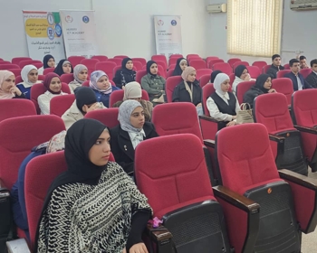
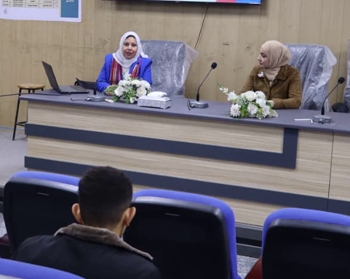
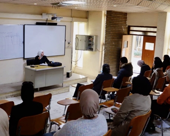
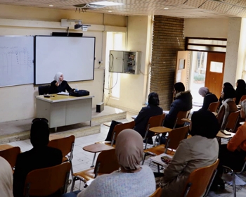
ناقش قسم علوم الحياة في كلية التربية للعلوم الصرفة (ابن الهيثم) رسالة ماجستير الموسومة ” التخليق الحيوي لجسيمات الفضة النانوية باستخدام المستخلصات النباتية ودراسة تأثيرها على البكتريا المعزولة من المرضى المصابين بالتهاب المجاري البولية ” للطالبة ” هند ناجي عبيد ” التي انجزتها باشراف التدريسي في القسم ” أ.م.د. عصام جاسم خريبط ” ونوقشت من قبل اعضاء لجنة المناقشة المدرج اسمائهم في ما يأتي :
أ.د. رنا مجاهد عبد الله – رئيسا
أ.م.د. محمد عباس فياض – عضوا
أ.م.د. داليا باسل حنا – عضوا
أ.م.د. عصام جاسم خربيط – عضوا و مشرفا
ويهدف البحث الى :
-
جمع عينات البول وتشخيص العزلات البكتيرية.
-
الكشف عن حساسية العزلات البكتيرية لاصناف مختلفة من المضادات الحيوية باستخدام Vitek 2 compact system .
-
تحضير جسيمات الفضة النانوية باستخدام المستخلصات النباتية.
-
توصيف جسيمات الفضة النانوية باستخدام عدة تقنيات تحليلية.
-
اختبار نشاط جسيمات الفضة النانوية الحيوية في التطبيقات الطبية.
وتم في هذا البحث جمع مائة وستين عينة ادرار من المرضى المصابين بخمج المسالك البولية من بعض مستشفيات بغداد شملت مستشفى غازي الحريري للجراحات التخصصية، المختبرات التعليمية بمدينة الطب، مستشفى الاطفال المركزي، مستشفى الكرخ العام ومستشفى مدينة الامامين الكاظمين خلال الفترة من25/7/2019 الى 22/10/2020.
تم استنبات العينات على اوساط زرعية مختلفة تضمنت Eosin methylene blue) MacConkey agar, Blood agar, CHROMagar orientation) للتعرف على الخصائص الزرعية. شخصت العزلات البكتيرية ايضا بالاعتماد على الاختبارات الكيموحيوية. تم اجراء التشخيص التأكيدي للعزلات بواسطة جهاز الفايتك Vitek 2 compact system. أظهرت نتائج التشخيص انه تم الحصول على 66 عزلة منها40 (61%) تعود الى Escherichia coli ،16(24%)عزلة تعود الى Klebsiella pneumoniae، 5 عزلات (7.5%) عزلة Proteus mirabilis، عزلة2 (3%) Enterobacter spp.، 2 عزلة (3%) Pseudomonas aeruginosa، 1عزلة (1.5%) Staphylococcus aureus .
اظهرت نتائج اختبار الحساسية تجاه 14 مضاد حيوي بواسطة جهاز الفايتك ان اعلى نسبة مقاومة سجلت في عزلات E. coli تجاه مضاد trimethoprim/sulfamethoxazole بواقع 36 عزلة وبنسبة (90%) يليه مضاد ampicillin بواقع 34 عزلة وبنسبة (85%). في حين اظهرت جميع عزلات K. pneumoniae مقاومة لمضاد ampicillin بواقع 16 عزلة بنسبة (100%) تليها 14 عزلة (87.5%) مقاومة لمضاد ampicillin/sulbactam. أظهرت انماط المقاومة للمضادات الحيوية ان 30 (75%) من عزلات E. coli ذات مقاومة متعددة، بينما اظهرت 15عزلة من K. pneumoniae وبنسبة (97.3%) مقاومة متعددة للمضادات الحيوية. تم اجراء الكشف المظهري عن العزلات المنتجة لانزيمات البيتالاكتميز Extended Spectrum β-lactamase. اشارت النتائج الى ان 17عزلة (42.5%) من E. coli و5 عزلات (31.2%) K. pneumoniae منتجة ل ESBLs على التوالي.
أشارت نتائج الدراسة الحالية ان جميع عزلات E. coli, K. pneumoniae ليس لها القدرة على انتاج الهيمولايسين عند زراعتها على وسط اجار الدم. ابدت 32 عزلة من E. coli القابلية على تكوين الغشاء الحيوي وبنسبة 80% بطريقة Microtiter plate. من جانب اخر اظهرت النتائج ان جميع عزلات K. pneumoniae 16 (100%) منتجة للغشاء الحيوي.
تم استعمال المستخلص المائي لأوراق نبات الاوكالبتوس Eucalyptus camaldulensis كعامل اختزال لنترات الفضة في التخليق الحيوي لجسيمات الفضة النانوية. دل ظهور اللون البني الغامق على نجاح عملية التخليق الحيوي، وكانت الظروف الاولية في عملية التخليق الحيوي عند 1 ملي مولار، نسبة المستخلص النباتي الى محلول نترات الفضة 20:80 v/v))، PH 6.7، وقت التفاعل 25 دقيقة وعند 60 درجة مئوية. بينما اشارت النتائج الى ان الظروف المثلى للتخليق الحيوي لجسيمات الفضة النانوية كانت 2 ملي مولار، 20 دقيقة وعند 80 درجة مئوية.
تم توصيف جسيمات الفضة النانوية بواسطة مقياس الطيف الضوئي المرئي للأشعة فوق البنفسجية، المجهر الالكتروني الماسح، جهاز حيود الاشعة السينية ومطيافية فيوريه للأشعة تحت الحمراء. أشارت نتائج التوصيف الى تكون جسيمات الفضة النانوية عند الطول الموجي 421 نانومتر، اما متوسط حجم الجسيمات فقد تراوح بين 22.33 الى 74.43 نانومتر والحجم البلوري 20 نانومتر.
تمت دراسة التركيز المثبط الادنى والتركيز القاتل لجسيمات الفضة النانوية ضد العزلات ذات المقاومة المتعددة للمضادات الحيوية من E. coli, K. pneumoniae, S. aureus بتراكيز تراوحت من 500 الى 31.25 ميكروغرام/مل. أظهرت النتائج ان قيم MIC, MBC قد تراوحت من (62.5 الى 250 ميكروغرام/مل) ومن (125 الى 500 ميكروغرام/مل) على التوالي.
اظهرت نتائج دراسة تأثير جسيمات الفضة النانوية على تكوين الاغشية الحيوية على العزلات البكتيرية قيد الدراسة ان الفضة النانوية قد ثبطت الغشاء الحيوي المتكون بتركيز 62.5 ميكروغرام/مل وان النسبة المئوية للتثبيط قد زادت بزيادة التركيز الى 125 ميكروغرام/مل. كما اشارت النتائج الى ان الاغشية الحيوية المتكونة بواسطة K. pneumoniae اقل حساسية لجسيمات الفضة النانوية.
بينت نتائج التأثير التأزري لجسيمات الفضة النانوية مع مضاد ampicillin باستعمال طريقة الانتشار بالحفر ضد العزلات ذات المقاومة المتعددة من E. coli, K. pneumoniae, S. aureus ظهور مناطق تثبيط، أعلى منطقة تثبيط قد لوحظت في العزلة E14 (22 ملم). بينما لم يظهر مضاد ampicillin لوحده فعالية في تثبيط نمو العزلات البكتيرية.
كما اشارت النتائج الى ان دقائق الفضة النانوية المخلقة حيويا امتلكت نشاطا كبيرا في كسح الجذور الحرة بطريقة DPPH. اظهرت الفعالية المضادة للأكسدة نسبة 71.30% عند التركيز 400 ميكروغرام/مل. كما اظهرت قيم IC50 92.83، 59.56 ميكروغرام/ مل لحمض الأسكوربيك و الفضة النانوية على التوالي. تم فحص السمية الخلوية لدقائق الفضة النانوية على خط خلايا سرطان الثدي MCF-7 breast cancer cell lines وWRL68. دلت النتائج ان لدقائق الفضة النانوية تأثير سام على الخلايا بتراكيز تراوحت من 400 الى12.5 ميكروغرام/مل، كما لوحظ انخفاض حيوية الخلايا الى 48.23% عند التركيز 400 ميكروغرام/مل. بينما اظهرت النتائج ان دقائق الفضة النانوية كان لها تأثير سمي متوسط الى ضعيف على خط خلايا WRL68 normal cell line. كما لوحظ ان قيم IC50 MCF-7 ,WRL68 cell lines كانت 134.9، 66.32 ميكروغرام/مل على التوالي.
وخلصت الطالبة الى التوصيات:
1- إجراء المزيد من الدراسات حول مقاومة البكتيريا للمضادات الحيوية للحد من انتشارها وتطورها.
2- دراسة التخليق الحيوي لجسيمات الفضة النانوية باستخدام أجزاء أخرى من نبات Eucalyptus camaldulensis مثل الجذر والساق والزهور واللحاء.
3. دراسة المركبات الكيميائية النباتية لمستخلص أوراق E. camaldulensis يمكن استخدامها في التطبيقات العلاجية.
4- دراسة تأثير جسيمات الفضة النانوية المُصنَّعة حيوياً على عوامل الضراوة المختلفة للبكتريا المرضية.
5- دراسة سمية جسيمات الفضة النانوية الحيوية داخل الاجسام الحية باستخدام الحيوانات المختبرية.
Biosynthesis of silver nanoparticles using plant extract and study their effect on bacteria isolated from patients of urinary tract infection.
By Hind N.Obaid
Supervised by Assist.Prof.Dr. Essam Jasim
The objectives of study
.Collection of urine samples and identification of bacterial isolates
.Detection of antibacterial susceptibility of bacterial isolates against different classes of antibiotics using Vitek2 compact system.
.Preparation of silver nanoparticles using plant extract.
.Characterization of silver nanoparticles using several analytic techniques.
.Experience the activity of biogenic silver nanoparticles in medical application.
Summary
One hundred and sixty of urine samples were collected from some hospitals in Baghdad included (Ghazi Al-Hariri hospital for surgical specialist, Teaching laboratories of Medical City, Teaching Central Children hospital, Al-Karkh hospital and Al-Imamein Al-Kadhimein City hospital) during the period from 25/7/2019 to 22/10/2020.
The samples were cultured on different media (MacConkey agar, Eosin methylene blue, Blood agar and CHROMagar orientation) to identify the morphological characterization. Then, the isolates were subjected to conventional biochemical methods. The confirmed identification was performed using Vitek 2 compact system. After diagnostic methods, the results showed that 66 isolates were obtained 40 (61%) isolates belong to Escherichia coli, 16 (24%) isolates for Klebsiella pneumoniae, 5 (7.5%) isolates proteus mirabilis, 2 (3%) isolates Enterobacter spp., 2(3%) isolates Pseudomonas aeruginosa and 1 (1.5%) Staphylococcus aureus.
The sensitivity against 14 antimicrobial agents was demonstrated that isolates of E. coli were high resistance to trimethoprim/sulfamethoxazole 36 (90%) and ampicillin 34 (85%). While the isolates of K. pneumoniae displayed 16 (100%) resistance to ampicillin followed by 14 (87.5%) isolates resistance to ampicillin/sulbactam. On the other hand, the most effective antimicrobials for both isolates were amikacin and carbapenem antibiotics. The antibiotic resistance patterns of E. coli isolates exhibited 30 (75%) as multidrug resistance, whereas multidrug resistance K. pneumoniae isolates showed higher percentage 15 (97.3%). In addition, phenotypically Extended Spectrum β-lactamases producer isolates was detected. The results showed that 17 (42.5%) and 5 (31.2%) of E. coli and K. pneumoniae respectively were positive for ESBLs.
The results in this study revealed that both E. coli and K. pneumoniae isolates have no ability for hemolysin production when cultured on blood agar media. Biofilm formation was determined using microtiter plate method; the results demonstrate that 32 (80%) of E. coli isolates were biofilm producers, while all K. pneumoniae isolates 16 (100%) showed ability for biofilm production.
aqueous leaf extract of Eucalyptus camaldulensis was used as reducing agent for silver nitrate in biosynthesis of silver nanoparticles. The appearance of dark brown yellow indicated the success of biosynthesis process at the initial conditions (1mM of AgNO3, PH 6.7, plant extract to AgNO3 was 20:80 v/v, time reaction was 25 minutes and temperature was 60 ºC). The optimal conditions for biosynthesis of silver nanoparticles were recorded at 2mM of AgNO3, 20 minutes and 80 ºC.
The synthesis of silver nanoparticles was confirmed b UV-Visible spectrophotometer, scanning electron microscopy (SEM), X-ray diffraction (XRD) and Fourier transform infrared spectroscopy (FTIR). The results of characterization indicated the formation of silver nanoparticles. The λmax of UV-visible were recorded at wavelength 421 nm. The average particle size range from 22.33 to 74.43 nm and crystalline size was 20 nm.
Minimum inhibitory concentration and minimum bactericidal concentration were examined against multidrug resistance isolates of E. coli, K. pneumoniae and Staphylococcus aureus at the concentration ranged from 500 to 31.25 µg/ml. The results showed that MIC and MBC ranging from (62.5 to 250 µg/ml) and (125 to 500 µg/ml) respectively.
The effect of silver nanoparticles on biofilm formation was detected among the isolates of urinary tract infection. The results indicated that silver nanoparticles inhibited biofilm at the concentration of 62.5 µg/ml, but the percentage of inhibition was increased with increased concentration to 125 µg/ml. K. pneumoniae biofilm was found to be less susceptible than E. coli at the same concentrations.
As for synergistic effect, the silver nanoparticles were combined with ampicillin using well diffusion method against multidrug resistance isolates of E. coli, K. pneumoniae and S. aureus. The results demonstrated that the higher inhibition zone was observed in E. coli (E14 isolate) of 22 mm and the lowest inhibition (15 mm) was recorded against S. aureus, while ampicillin alone do not.
Silver nanoparticles exhibited significant scavenging activity of free radical by DPPH method and the maximum percentage of antioxidant activity showed 71.30% at 400 µg/ml with IC50 59.56 and 92.83µg/ml for ascorbic acid and AgNPs respectively.
The cytotoxicity of silver nanoparticles on MCF-7 breast cancer and WRL68 cell lines was evaluated using MTT assay. The results revealed that silver nanoparticles have cytotoxic effect at the concentrations ranging from 400 µg/ml to 12.5 µg/ml. The viability of MCF-7 breast cancer cell line was decreased to 48.23% at 400 µg/ml with IC50 66.32 µg/ml. Whereas, the effect of AgNPs on WRL68 normal cell line showed moderate to weak toxicity and exhibited IC50 of 134.9 µg/ml.
Recommendations
.Conduct more studies on bacterial resistance to antibiotics for limiting its spread and development.
.Study the biosynthesis of silver nanoparticles using other parts of Eucalyptus camaldulensis plant such as root, stem, flower and bark.
Study the phytochemical compounds of E. camaldulensis leaf extract can be use in therapeutic applications.
.Study the action of biosynthesized silver nanoparticles on different virulence factors of pathogens.
.Study the toxicity of biogenic silver nanoparticles in vivo on model animals.

