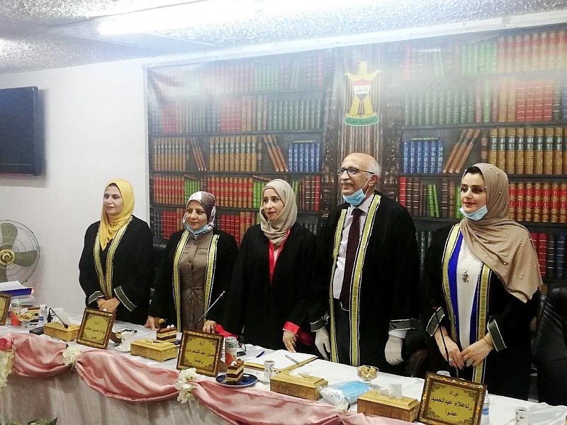
ناقش قسم علوم الحياة في كلية التربية للعلوم الصرفة (ابن الهيثم) رسالة الماجستير الموسومة ( دراسة شكليائية ونسجية مقارنة للسان في السنجاب القوقازي Sciurus anomalus وبيز ابن عُرس Herpestes javanicus ) للطالبة ( مروه خليل ابراهيم ) التي انجزتها تحت اشراف التدريسية في القسم ( أ.م.د. إيمان سامي أحمد ) و نوقشت من قبل لجنة المناقشة التي تألفت من الاعضاء المبينة اسمائهم فيما يأتي :
-
أ.د. حسين عبد المنعم داود (رئيسا)
-
أ.م.د. بيداء حسين مطلك (عضوا)
-
م.د. رنا علاء عبد الحميد (عضوا)
-
أ.م.د. ايمان سامي احمد (عضوا و مشرفا)

هدفت الدراسة الحالية للتعرف على الوصف الشكليائي والتركيب النسجي للسان في نوعين من الثدييات العراقية متمثلة بالسنجاب القوقازي Sciurus anomalus وبيز ابن عُرس Herpestes javanicus والمقارنة بينهما ، واظهرت نتائج الدراسة ما يأتي:-
الوصف الشكليائي:
يتموضع اللسان في كلا حيوانين موضوع الدراسة الحالية في التجويف الفمي اذ يرتبط بقاعه بوساطة اللجام الذي يبدو قصيراً في السنجاب القوقازي بينما يكون طويلاً وواسعاً في بيز ابن عُرس الذي يمتد منه تركيباً قضيبياً يدعى ليزا Lyssa . يبدو اللسان في السنجاب القوقازي متطاولاً وذو نهاية قمية مستديرة وسميكة ويتميز بوجود تخصرٍ عند الثلث الاول منه فيما يكون اللسان في بيز ابن عُرس اكثر طولاً وذو قمة مستديرة ونحيفة . ينفرد لسان السنجاب القوقازي بوجود ارتفاعٍ ضئيلٍ يتموضع عند الجزء الوسطي من السطح الظهري يمثل بروزاً لسانياً اثرياً في حين يتميز لسان بيز ابن عُرس بوجود انخفاض بسيط يدعى بالحفرة اللسانية التي تأخذ شكلاً دائرياً تتموضع عند الجزء القحفي من الجسم اللساني .
يمتلك حيوانا الدراسة الحالية نوعين من الحليمات اللسانية ، تتمثلان بحليمات ميكانيكية واخرى ذوقية تضم الاولى حليمات خيطية فقط في السنجاب القوقازي في حين تضم في بيز ابن عُرس على حليمات خيطية واسطوانية اما الحليمات الثانية فتنضوي تحتها حليمات فطرية ومسوّرة وورقية في السنجاب القوقازي بينما تتضمن في بيز ابن عُرس على حليمات فطرية ومسوّرة فقط . تنتشر الحليمات الخيطية على السطح الظهري والسطحيين الجانبيين للسان في كلا الحيوانين بينما ينفرد السنجاب القوقازي بوجود اعداد من الحليمات الخيطية تتموضع عند السطح البطني للقمة اللسانية في حين تنتشر ذات الحليمات في بيز ابن عُرس عند جانبي السطح البطني للجسم اللساني. يتميز لسان بيز ابن عُرس بامتلاكه لحليمات اسطوانية تتواجد في الحفرة اللسانية ، اما فيما يخص الحليمات الفطرية فانها تتناثر بين الحليمات الخيطية على السطح الظهري والسطحين الجانبيين لكلا حيواني الدراسة الحالية الا ان هذه الحليمات تتناثر اعداد منها عند السطح البطني للقمة اللسانية فضلاً عن انتشارها بين الحليمات المسوّرة للسان السنجاب القوقازي فقط بينما ينفرد لسان بيز ابن عُرس بوجود حليمات فطرية عملاقة تتموضع على جانبي القمة اللسانية . تتميز الحليمات المسوّرة في كلا الحيوانيين موضوع الدراسة كونها مؤلفة من ثلاث حليمات تترتب بشكل V)) مقلوب وان لكل حليمة جزء وسطي مستدير محاط بخندق دائري وهذا بدوره يطوق بوسادة لكنه تبين ان الحليمات المسوّرة لدى السنجاب القوقازي تبرز عن مستوى سطح اللسان وتظهر الوسادة فيها نحيفة في حين تبدو الحليمات المسوّرة في بيز ابن عُرس غائرة ضمن وسادة سميكة ذات شكل شبيه بالدمعة باستثناء الحليمة الثالثة التي تظهر فيها الوسادة كاسية الشكل .
تميز لسان السنجاب القوقازي بامتلاكه على حليمات ورقية تتموضع على جانبي الجزء الذيلي من الجذر اللساني التي تبدو بشكل طيات متوازية تفصلها اخاديد ، تترتب حافاتها الحرة بزاوية مائلة لتتجه نحو الحليمات المسوّرة .
التركيب النسجي:
يتألف اللسان نسجياً في السنجاب القوقازي من ثلاث غلالات متمثلة بالغلالة المخاطية ، والغلالة تحت المخاطية – الصفيحة الاصيلة ، والغلالة العضلية اما اللسان في بيز ابن عُرس فيتالف من الغلالة المخاطية والغلالة تحت المخاطية والغلالة العضلية . كما تبين ان الغلالة المخاطية في السنجاب القوقازي مؤلفة من البطانة الظهارية فقط التي تتكون من نسيج ظهاري حرشفي مطبق متقرن في حين تبدو الغلالة المخاطية في بيز ابن عُرس مؤلفة من البطانة الظهارية والصفيحة الاصيلة ، تتكون الاولى كما في السنجاب القوقازي من نسيج ظهاري حرشفي مطبق متقرن اما الصفيحة الاصيلة فتتالف من نسيج ضام مفكك ، وتتكون الغلالة تحت المخاطية –الصفيحة الاصيلة في السنجاب القوقازي من نسيج ضام مفكك ،بينما تبدو الغلالة تحت المخاطية في بيز ابن عُرس مكونه من نسيج ضام كثيف . اما بخصوص الغلالة العضلية في كلا الحيوانين فانها مؤلفة من الياف عضلية هيكلية تترتب عموماً بشكل حزم وباتجاهات مختلفة ، طويلة وعريضة ومائلة ، كما انها تتباين في ترتيب اتجاهاتها مابين لسان الحيوانين ومابين مناطق لسان الحيوان الواحد .
تتالف الحليمات اللسانية عموماً في كلا الحيوانين من البطانة الظهارية واللب ، تتمثل البطانة الظهارية بنسيج ظهاري حرشفي مطبق متقرناً بدرجات متفاوته في الحليمات الميكانيكية بينما يبدو في الغالب غير متقرناً في الحليمات الذوقية التي تتخلله اعداد متباينة من البراعم الذوقية ، اما اللب فيتالف من نسيج ضام مفكك الذي يمثل امتداداً للغلالة تحت المخاطية –الصفيحة الاصيلة في السنجاب القوقازي فيما يعد امتدادا للصفيحة الاصيلة في بيز ابن عُرس . تتموضع الغدد اللسانية في كلا الحيوانين على جانبي الجذر اللساني ويحتل جزءها الاكبر الغلالة العضلية الا انها تبدا من الغلالة تحت المخاطية – الصفيحة الاصيلة في السنجاب القوقازي ومن الغلالة تحت المخاطية في بيز ابن عُرس وتتضمن نوعين من الغدد ، يتمثل الاول بالغدد المصلية (فون ايبنر Von –Ebner ) التي تفتح اقنيتها في خندق الحليمات المسوّرة فيما يتمثل النوع الثاني بالغدد المخاطية والمخاطية المصلية (الغدد المختلطة) التي تدعى غدد ويبر Weber وتفتح اقنيتها على سطح الجذر اللساني ويتغلغل هذا النوع من الغدد ليصل الى لب اللسان في السنجاب القوقازي ،ان نتائج الكشف عن نوع افراز المخاطين لغدد ويبر باستعمال الملونين شف الحامض الدوري والاليشين الازرق (pH:2.5 ) بينت ان استجابته لملون الاليشين تبدو اكثر في السنجاب القوقازي الا انها ظهرت على العكس من ذلك في بيز ابن عُرس اذ تبدو استجابته لملون شف الحامض الدوري اكثر .
وخلصت الطالبة الى التوصيات الاتية :
افرزت نتائج الدراسة الحالية مجموعة من النقاط تحتاج الى تسليط الضوء عليها من خلال اجراء المزيد من الدراسة والتقصي بغية الوصول الى الحقائق العلمية وبناءً الى ذلك نوصي بما يأتي:-
-
دراسة اللسان على مستوى المجهر الالكتروني الماسح Scanning electron microscope للحيوانين موضوع الدراسة لتعذر دراستها من قبل الباحث لاسباب فنية ، على ان يكون ذلك موضوعاً لمشروع دراسة مستقبلية.
-
احتساب سمك الغلالات لجميع الحليمات اللسانية في كلا حيواني الدراسة الحالية والمقارنة بينهما.
-
اجراء دراسات شكليائية ونسجية مقارنة للسان في انواع اخرى من الفقريات البرية العراقية تبعاً لنوع التغذية بُغية وضع اساس علمي للدراسات الاخرى التي لم تنل الاهتمام العلمي الكافي.
-
اجراء دراسة نسجية وجنينية للسان في الفقريات العراقية.
-
استعمال تقنيات حديثة في وسم طبقات الحليمات اللسانية ب CD marker والتقنية الكيميائية النسجية المناعية Immunohistochemistry في تلوين المقاطع النسجية.
Comparative Morphological and Histological Study of The Tongue In Sciurus anomalus and Herpestes javanicus
By Marwa .K.Ibrahim
Supervised by Asst.prpf.Dr.Eman.S.Ahmed
Summary
The current study is aimed to investigate the morphological description and the histological structure of tongue in two species of Iraqi mammals caucasian squirrel ( Sciurus anomalus ) and mongoose ( Herpestes javanicus ) and compared with them. The results of this study showed the following:
Morphological description:
The tongue is situated in both animals of the preseut study subject in oral cavity, which is connected with its bottom by frenulum.The frenulum tends to be short in the caucasian squirrel, while it is long and wide in mongoose, which extends from it a rod-like structure called lyssa. The tongue in the caucasian squirrel seems long and has a round and thick tip and is characterized by the presence of a narrowing at the first third of it, while the tongue in the mongoose is more long and has a round thin tip .
The caucasian squirrel tongue is unique in the presence of a small height positioned at the middle part of the dorsal surface represents an lingual prominence while the tongue of mongoose is characterized by a small reduction called the lingual fossa that takes a circular shape positioned at the cranial part of the lingual body. The two animals of the current study have two types of lingual papillae, consisting of mechanical and gustatory papillae. The first type includes only filiform papillae in caucasian squirrel,while includes filiform and cylindrical papillae in mongoose. As for the second papillae is included the fungiform, circumvallate and foliate in caucasian squirrel, whilst in mongoose is included only fungiform, circumvallate papillae.
The filiform papillae is spread on the dorsal and two laterals surfaces of the tongue in both animals, but the caucasian squirrel is unique in the presence of a number of filiform papillae located at the ventral surface of the lingual apex, while in mongoose it is spread on the two laterals of ventral surface of lingual body. The tongue of mongoose is characterized by its possession of cylindrical papillae located in the lingual fossa . As for the fungiform papillae, they are scattered between the filiform papillae on the dorsal surface and the two laterals surfaces of both animals in the current study. These papillae are scattered numbers of them at the ventral surface of the lingual apex as well as spread among the circumvallate papillae of the caucasian squirrel’s tongue only, while mongoose’s tongue is unique in the presence of giant fungiform papillae placed on both sides of the lingual apex.
The circumvallate papillae in both animals of this study are characterized by the fact that it consists of three papillae arranged as form of the inverted letter (V) . Each papilla has a round middle part surrounded by a circular groove and this in turn surrounds a pad . It shows that the circumvallate papillae in caucasian squirrel emerged from the level of the surface of the tongue as well as the pad is thin, while the circumvallate papillae in mongoose, appears confluent within thick pad and form of tear -like except for the third papilla, in which the pad appears to be a calyx form.
The tongue of the caucasian squirrel is characterized by its possession of the foliate papillae placed on the two laterals of the caudal part of the lingual root, which appears in parallel folds separated by grooves, its free edges at oblique angle towards the circumvallate papillae.
Histological structure
The tongue of the caucasian squirrel is formed of three tunicae represented by mucosa, submucosa- lamina propria and muscularis, while the tongue is formed in mongoose of tunicae mucosa, submucosa and muscularis. It is also found that the tunica mucosa in the caucasian squirrel consist of only lining epithelium, which consist of keratinized stratified squamous epithelial tissue, while the tunica mucosa in mongoose appears to be composed of the lining epithelium and the lamina propria. The first, as in the caucasian squirrel consists of keratinized stratified squamous epithelial tissue, and the lamina propria is formed of loose connective tissue, The submucosa-lamina propria consists of loose connective tissue in caucasian squirrel ,while the tunica submucosa in mongoose appears to be formed of dense connective tissue. As for the tunica muscularis in both animals, it is composed of skeletal muscular fibers that generally arranged as form of bundles and in different directions, longitudinal, transverse and oblique. As they vary in their directions between the tongue of the two animals and between the areas of the tongue in the one animal. The lingual papillae is generally composed in both animals of lining epithelium and core, the lining epithelium is represented by keratinized stratified squamous epithelial tissue which is varying degrees in mechanical papillae but it appears mostly non-keratinized in the gustatory papillae that interspersed with a variety of taste buds. Whereas the core is formed of a loose connective tissue, which represent an extension of the submucosa- lamina propria in the caucasian squirrel, whilst is an extension of the lamina propria in the mongoose.
The lingual glands in both animals are positioned on both sides of the lingual root and it’s mostly occupied by the tunica muscularis, but they start from the tunica submucosa– lamina propria in the caucasian squirrel and from the tunica submucosa in the mongoose and includes two types of glands, the first being the serous glands (von Ebner), which opens its ducts in the groove of the circumvallate papillae, the second type is the mucosal and seromucosal glands (mixed glands) called Weber glands which opens its ducts on the surface of the lingual root and penetrates this type of glands to the core of the tongue in caucasian squirrel . The results of the detection about the type of secretion of mucin in weber gland using Schiff’s periodic acid stain and Alcian blue (pH:2.5) showed that its response to the Alcian blue stain appeared more in the caucasian squirrel, but on the contrary in the mongoose, as its response to the Schiff’s periodic acid stain seems to be more.


