

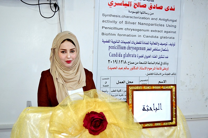
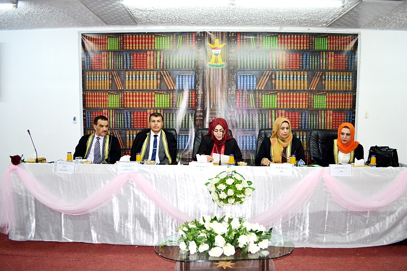
ناقش قسم علوم الحياة في كلية التربية للعلوم الصرفة (ابن الهيثم) رسالة الماجستير الموسومة ( توليف، توصيف والفعالية المضادة للفطريات لجسيمات النانوية الفضية باستعمال مستخلص الفطر Penicillium chrysogenum ضد تشكيل الغشاء الحيوي لخميرة Candida glabrata ) للطالبة (ندى صادق صالح ) التي انجزتها تحت اشراف التدريسي في القسم ( أ.م.د. ثامر عبد الشهيد محسن ) .
هدفت الدراسة الى استعمال الفطرPenicillium chrysogenum لتخليق جسيمات النانوية الفضية متناهية الصغر وتأثيرها ضد الغشاء الحيوي Biofilm للفطر Candida glabrata الممرض للانسان وقد تضمنت الدراسة المحاور الأتية:
-
عزل وتشخيص الخميرة Candida glabrata وتحديد الغشاء الحيوي لها.
2- بناء وتوصيف جسيمات النانوية الفضية باستعمال الفطرPenicillium chrysogenum وبأنواع مختلفة من المجاهر.
3- تحديد أقل تركيز مثبط (The Minimum Inhibitory Concentration (MIC لجسيمات النانوية الفضية ضد خميرة Candida glabrata .
4- دراسة التأثير التآزري لجسيمات النانوية الفضية للفطر Penicillium chrysogenum والعامل المضاد الفطري ضد الغشاء الحيوي لخميرة Candida glabrata.
5- دراسة التأثيرات النانوية الفضية على التغيرات النسيجية للفئران المصابة بالخميرة Candida glabrata.
وأظهر توزيع المرضى المصابين بداء مبيضات الدم والفم إن الفئة العمرية 50-65 سجلت أعلى إصابة بداء المبيضات في النساء والرجال المصابين بمرض سرطان الدم وبنسبة 50% و 37.9% على التوالي مقارنةً باقل إصابة بداء المبيضات للفئة العمرية دون 17 سنة في النساء والرجال وبنسبة 8.8% و13.5% على التوالي ، في حين كانت الفئة العمرية التي تتراوح بين 5-8 سنوات مرتفعة إذ وصلت الى 18 إصابة بداء المبيضات الفموي وبنسبة 42% للاطفال المصابين بسرطان الدم مقارنة مع الفئة العمرية 8-10 سنة والتي كانت أقل الاصابات إذ بلغت 9 حالات وبنسبة 21% وبفروق معنوية بين معاملات النساء والرجال بينما لم تسجل فروق معنوية بين الاطفال . سجلت أعلى الاصابات بخميرة Candida glabrata إذ بلغت 59 عزلة بين النساء والرجال المصابين بسرطان الدم في حين كانت خميرة C.kefyer اقل الاصابات وبعزلة واحدة فقط، كانت خميرة C. albicans أكثر العزلات الفموية المعزولة من الاطفال المصابين بسرطان الدم إذ بلغت 21 عزلة وتلتها خميرة C.glabrataبلغت 13 عزلة أما خميرة C. kefyer فكانت عزلتين فقط . أظهرت خميرة C.glabrataأعلى تكوين للغشاء الحيوي Biofilm فكانت 48 عزلة توزعت بين 21 إنتاجاً قوياً و 27 إنتاجاً ضعيفاً للغشاء أما أقل الخمائر إنتاجاً للغشاء كانت خميرة C. tropicalis إذ وصلت الى 6 عزلات وتوزعت بين 4 إنتاجات قوية وانتاجين ضعيفين ووجدت فروق معنوية بين الغشاء الحيوي وأنواع المبيضات المختلفة. لوحظ تغير اللون بعد مزج الكتلة الحيوية لفطر Penicillium chrysogenum مع محلول نترات الفضة من الاصفر الى البني عند دراسة تصنيع جسيمات النانوية الفضة بفعل وجود البلازمونات السطحية ، لتوصيف الجسيمات استعمل مجهر القوة الذرية AFM ووجد ان قيمة متوسط الجذر التربيعي 2.12 نانومتر وأن معدل الخشونة السطحية للغشاء 1.54 نانومتر وهذه القيمة تعد دليلا لخشونة السطح، ظهرت اعلى قمة بطول موجي 455 نانومتر بسبب تواجد البلازمونات السطحية وقمة اخرى بطول موجي 243 نانومتر وهذه تدل على وجود بقايا التايروسين Tyrosine والتربتوفان Tryptophan الموجودة في البروتين التي تحررت من الفطر عند استعمال مطياف الاشعة فوق البنفسجية، بينت نتائج المجهر الالكتروني الماسح SEM وجود حبيبات كروية الشكل متفاوتة الاحجام وموزعة بصورة منتظمة وبعض التجمعات والتكتلات اثناء النمو البلوري وذات احجام نانوية. سجلت نتائج تحديد التركيز الادنى المثبط لدقائق نانوية الفضة بأن جميع التراكيز المستعملة كان لها فعالية في تثبيط خميرة C.glabrata وكان افضلها التركيز 20 ملي مولاري الذي سجل اعلى منطقة تثبيط له بلغت 13 مليمتر وكان اقلها التركيز 5 ملي مولاري الذي اظهر اقل منطقة تثبيط اذ بلغت 7 مليمتر وكانت الفروق معنوية جداً. أظهرت خميرة C.glabrata قابليةعلى الالتصاق بالخلايا الطلائية عند عدم وجود جسيمات نانوية الفضة والمضاد الفطري، اما عند اضافة جسيمات الفضة النانوية فان الخميرة تفقد قابليتها على الالتصاق، في حين ان إضافة المضاد الفطري الفلوكونزول تسبب في تقليل التصاق الخميرة بالخلايا الطلائية أماعند جمع الجسيمات النانوية الفضية والمضاد الفطري فلوكونوزول فان خميرة C.glabrata تفقد قابليتها على الالتصاق نهائياً وتسبب في موتها. بينت نتائج التشريح النسجي لكبد الفئران إن مجموعة السيطرة من حيث شكل الخلايا وفصيصات وجيوب الكبد والوريد المركزي كانت طبيعية مع عدم وجود أية أضرار ظاهرة أو تغيرات نسجية، أما المجموعة المصابة بخميرة C.glabrata فقد لوحظت تغيرات نسجية واضحة مثل توسع واحتقان في الوريد المركزي وكذلك احتقان الوريد البابي والخلايا ثنائية الانوية وترشح والتهاب الخلايا، وأظهرت نتائج المجموعة التي تم اعطاؤها جسيمات نانوية فضية زيادة الياف الانسجة الضامة وترشح الخلايا الملتهبة المزمنة ووجود نواة غامقة في خلايا الكبد وبداية ظهور فجوات السايتوبلازم، أما مجموعة الفئران المصابة بخميرة C.glabrata والمعالجة بالجسيمات النانوية الفضية والفلوكونزول معاً ظهور تحسن واضح إذ لوحظ توسع خفيف في الجيوب والوريد المركزي ووجود تسطح في الوريد وترشح للخلية احادية النواة. نواة خلايا الكبد حوصلية ومصبوغة باللون الغامق ووجود فجوات في السايتوبلازم مع وجود خلايا ثنائية النواة.
Synthesis, Characterization and Anti fungal activity of Silver Nano Particles Using Penicillium chrysogenum extract against Bio film formation in Candida glabrata
By Nada Sadiq Salih
Supervised by Asst.prof.Dr.Thamir A.A Muhsen
Summary
The distribution of patients with candidiasis and oral disease showed that the age group 50-65 years recorded the highest incidence of candidiasis in women and men with leukemia by 50% and 37.9 % respectively compared to the lowest incidence of candidiasis in the age group under 17 years in women and men 8.8% and 13.5%, respectively, while the age group between 5-8 years was high, reaching 18 cases of oral candidiasis and 42% of children with leukemia compared with 8-10 years, which was the least. It reached 9 cases (21%) and found very significant differences between the treatment of women and men,No significant differences were recorded among the children. The highest incidence of Candida glabrata was 59 isolates of women and men with leukemia, while C. kefyer was the least and only one isolate. C. glabrata was the most isolated from children with leukemia 13and C.kefyer was only 2.
-
glabrata showed the highest bio film formation, 48 isolates were distributed between 21 strong and 27 weak biofilm production, The least biofilm producing isolates were C.tropicalis, which reached 6 isolates and distributed between 4strong and 2weak,There were very significant differences between the biofilm and different Candida spp. The color change was observed after the biomass of Peniclliumchrysogenum was mixed with the solution of silver nitrate from yellow to brown due to the presence of surface plasmons, For particle characterization, an atomic force microscope was used and the mean square root value was 2.12 nm and the surface roughness rate of the membrane was 1.54 nm this value is a guide to surface roughness, the highest peak with a wavelength of 455 nm has emerged due to the presence of surface plasmons and another peak with a wavelength of 243 nm these indicate the presence of Tyrosine and Tryptophan residues in the protein released from the fungus. when using UV spectrometer the results of the scanning electron microscope showed spherical granules of varying sizes some clusters are regularly distributed during crystalline growth and of nanoscale sizes.
The results of determining the minimum inhibitory concentration of silver nanoparticles were recorded that all concentrations used were effective in inhibiting C.glabrata, The best concentration was 20 mM, which recorded the highest inhibition zone of 13 mm, the lowest concentration was 5 mM, which showed the lowest inhibition zone at 7 mm and the differences were very significant.C. glabrata was observed for adhesion to epithelial cells in the absence of silver nanoparticles and antifungal whereas when silver nanoparticles were added, yeast lost its ability to adhere, the addition of the anti-fungal fluconazole caused reducing the yeast adhesion to the epithelial cells, when collecting silver nanoparticles and fluconzole, the C.glabrata is lost to final adhesion and caused her death.Histopathological results of mice liver showed that control group display normal liver lobules, liver sinusoids ,central vein and normal shape hepatocytes with no visible damage or histological changes.
The group infected with Candida glabratahad obvious histological changes such as display dilated congested central vein , congestion portal vein, bi-nucleated cells are also seen and inflammatory cell infiltration.The results of a group that were given silver nano particles observed increasing amount of connective tissue fibers,chronic inflammatory cell infiltration, hepatocytes have darkly-stained nuclei,and vacuolated cytoplasm could be established. While the group of mice infected with the fungus Candida glabrata and treated with silver nanoparticles and fluconzole showed slightly dilated sinusoids, central vein with flat endothelial lining ,Mononuclear cellular infiltration,hepatocytes with vesicular nuclei and cytoplasm ,darkly-stained nuclei,less vacuolated cytoplasm and bi-nucleated cells